Autoimmune Hemolytic Anemia: Causes, Diagnosis, and Treatment Options

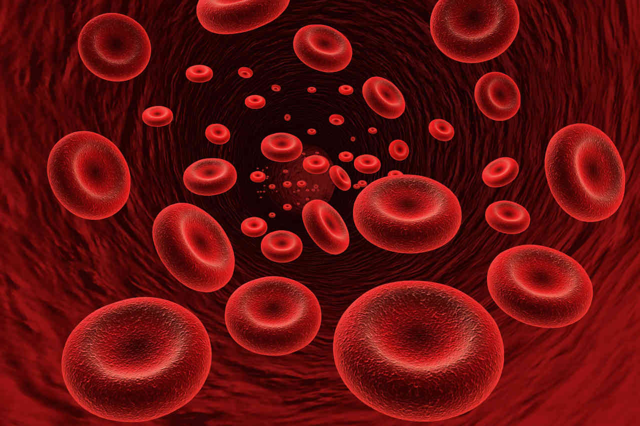
Autoimmune hemolytic anemia is a rare medical condition where the body’s own immune system attacks its red blood cells by mistake and destroys them. Red blood cells carry oxygen in your body. As a result, their shortage makes one feel tired or short of breath.

Speak to a Specialist About Copay Assistance

Schedule a Consultation

Normally, red blood cells have an average lifespan of 120 days. In autoimmune hemolytic anemia (AIHA), your body’s immune system mistakes red blood cells for unwanted substances, and it attacks the red blood cells and destroys them. As a result, your red blood cells die early. In severe cases, the red blood cells may exist for only a few days.

What Are the Types of Autoimmune Hemolytic Anemia (AIHA)?

Autoimmune hemolytic anemia (AIHA) is classified in 2 ways: Warm vs. cold and primary vs. secondary.

The warm and cold classification of AIHA depends on the antibody involved:

  • Warm AIHA: This type of anemia happens at or above normal body temperature. It involves IgG antibodies and is the most common type.
  • Cold AIHA: This type of anemia happens in the presence of IgM autoantibodies. This type of anemia happens when you are exposed to cold.

AIHA can also be classified as follows:

  • Primary AIHA: AIHA is classified as primary if it develops without any presence of an underlying condition.
  • Secondary AIHA: AIHA is classified as secondary if it is linked to an underlying condition like other autoimmune disorders, cancer, or viral infections.

Symptoms

Woman with headache as a symptom of autoimmune hemolytic anemia

Autoimmune hemolytic anemia (AIHA) is a condition where the body’s own immune system attacks and destroys red blood cells. AIHA can have many signs and symptoms. Some common symptoms of AIHA include:

  • Weakness
  • Difficulty breathing
  • Dark urine
  • Thinking and concentration difficulties
  • Fever
  • Rapid heartbeat
  • Jaundice (yellowing of skin or whites of eyes)
  • Headache
  • Muscle pains
  • A sore tongue

What Causes Autoimmune Hemolytic Anemia (AIHA)?

In approximately half of the cases, the exact cause of autoimmune hemolytic anemia (AIHA) is unknown. In other cases, links can be found between AIHA and some other conditions. There could be several factors and disease states that may contribute to the development of AIHA. These include:

Underlying Autoimmune Disorders: Several autoimmune diseases can contribute to the development of AIHA. Some of them include lupus, autoimmune thyroid disease, ulcerative colitis, and rheumatoid arthritis. In these cases, the immune system may produce antibodies that not only target the cells of the associated autoimmune disorder but also attack red blood cells.

Viral Infection: Some viral infections can cause AIHA. These include measles, hepatitis, mumps, epstein-barr virus, and rubella. However, once the infection is treated, the anemia goes away.

Genetics: Parents can pass the gene for the condition on to their children [2].

Medications: In rare situations, certain drugs can trigger immune system responses that result in AIHA. These drugs can include NSAIDS (nonsteroidal anti-inflammatory drugs), anti-cancer drugs, methyldopa, quinidine, and certain antibiotics [1].

Diagnosis

The diagnosis process of AIHA begins with examining the clinical history of the patient. A doctor will identify symptoms related to anemia. After that, the doctor will suggest some tests to confirm AIHA. These tests are:

  • Complete Blood Count (CBC): A CBC reveals a low red blood cell count (anemia) in AIHA. Also, it can show increased reticulocyte count, which indicates increased red blood cell production in the body, and the size of the red blood cells the body is making.
  • Peripheral Smear: In this test, a healthcare professional examines your blood sample under a microscope to see if your blood cells are being destroyed.
  • Coombs Test: This test detects the presence of antibodies that target red blood cells. This test can also tell if anyone has warm AIHA or cold AIHA.
  • Lactate Dehydrogenase (LDH): Lactate dehydrogenase is an enzyme found in red blood cells. When red blood cells are damaged, the LDH level increases.
  • Cold Agglutinin Titer: If your doctor suspects cold AIHA, he may suggest this test. This test shows the level of the antibody responsible for cold AIHA.

Once diagnosed, your doctor will tailor the treatment plan according to the type and severity of your AIHA and any underlying condition.

Treatment of Autoimmune Hemolytic Anemia (AIHA)

You do not need any treatment if your anemia is mild. It often passes without any treatment. However, in other cases, you may need medications, surgery, or a blood transfusion. 

The treatment of autoimmune hemolytic anemia (AIHA) begins with addressing any underlying condition first. If any medication, like penicillin, is causing your anemia, then you have to stop using that medicine. If you have any condition, like lupus or rheumatoid arthritis, that is causing your anemia, then your doctor will treat that condition first.

Here are some common treatment options for autoimmune hemolytic anemia (AIHA):

Medications: The first line of treatment for AIHA is a class of drugs called corticosteroids. They help weaken your body’s immune system and reduce the production of antibodies that target red blood cells. If corticosteroids don’t work, then your doctor may prescribe immunosuppressive medications. Doctors can also prescribe a drug called rituximab. This drug reduces the production of antibodies that target red blood cells [3].

Surgery: If medicines don’t work, then your doctor may recommend surgery to remove your spleen. Your spleen destroys the abnormal red blood cells, including those attached to antibodies. Therefore, the removal of the spleen can decrease your anemia. 

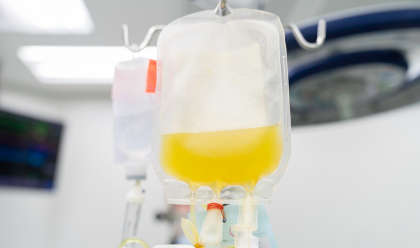
Blood Transfusion: In severe cases, you may need a blood transfusion.

Autoimmune Hemolytic Anemia: FAQs

Here are some common faqs about autoimmune hemolytic anemia:

How Can You Prevent Autoimmune Hemolytic Anemia (AIHA)?

It is not always possible to prevent autoimmune hemolytic anemia (AIHA). This is because, in many cases, we can not determine the exact cause of AIHA. However, if you take medicines that have a link to AIHA, then your doctor may monitor you closely. This will reduce your risk of developing this condition.

How Common Is Autoimmune Hemolytic Anemia?

Autoimmune hemolytic anemia (AIHA) is a rare autoimmune disorder. It only has an annual incidence of 1 – 3 cases per 100,000 people.

What Happens If You Do Not Treat Autoimmune Hemolytic Anemia (AIHA)?

For people with mild AIHA, no treatment is needed. However, treatment is necessary if you have serious anemia. If you do not treat the condition, it can cause heart problems. In severe cases, it can even cause heart failure or death [4].

Who Is Affected Most by Autoimmune Hemolytic Anemia (AIHA)?

Although this disease can happen at any age, people over the age of 40 are most likely to be affected by it. Additionally, females have a greater chance of developing this condition than men [5].

Can AIHA Affect Pregnancy?

During pregnancy, AIHA can cause severe life-threatening anemia in 40 – 50% of cases. It can also cause stillbirth. Also, 35 – 40% of all infants from mothers with AIHA may be born with severe postpartum hemolytic anemia [6].

How To Test for Guillain-Barre Syndrome

Routine tests for Guillain-Barre syndrome are cerebrospinal fluid (CSF) analysis, electromyography, and nerve conduction studies. Additional tests include an MRI (magnetic resonance imaging) or a CT (computed tomography) scan. 

Speak to a Specialist About Copay Assistance

(877) 778-0318

Diagnosing Guillain-Barre syndrome (GBS) can be challenging, especially during the earlier stages. GBS shares many signs and symptoms with other neurological disorders.

Moreover, the symptoms can vary from one person to another. 
Before ordering a specific test for Guillain-Barre syndrome, your doctor will assess your symptoms and medical history. If they suspect GBS, they may order lab tests to check your blood sugar, electrolytes, kidney function, and liver enzymes [1].

Though these tests are of little diagnostic value, they can help rule out other causes responsible for your symptoms, such as: 

  • Infections 
  • Metabolic disorders
  • Electrolyte dysfunctions

Specific Tests for Guillain-Barre Syndrome

Your doctor will likely recommend the following tests for Guillain-Barre syndrome to confirm the clinical diagnosis: 

Lumbar puncture

Also called a spinal tap, this test checks your CSF for abnormalities in protein levels or for signs of infections. 

Increased protein levels without an elevation in white blood cells is the characteristic feature of GBS [1]. Nonetheless, both may be normal during the first few weeks after disease onset.  

During a lumbar puncture, a healthcare provider inserts a tiny hollow needle into the spinal canal in the lower back to withdraw a sample of CSF. Potential risks of a lumbar puncture include transient numbness of the lower back, bleeding, or headache.

Electromyography

When your brain tells a muscle to move, the nerve signals travel in the form of electrical waves. Electromyography measures these electrical waves.   

During electromyography, tiny needles (electrodes) are inserted into the muscle. The electrodes measure electrical activity and display it on a monitor. Abnormal readings on the monitor may indicate muscle or nerve problems.

This test for Guillain-Barre syndrome can help support diagnosis in individuals with atypical signs and symptoms of other diseases. 

Nerve conduction studies

Nerve conduction studies measure how fast electrical signals move down the nerves. Abnormal results may indicate nerve damage. 
 

Other Tests for Guillain-Barre Syndrome

Imaging tests

MRI scan to rule out other conditions

Magnetic resonance imaging (MRI) and computed tomography (CT) scanning of the spine are not routinely used. However, they may help exclude:

  • Stroke
  • Spinal cord inflammation
  • Cancer involving the membranes surrounding your brain and spinal cord
  • Nerve root compression
  • Infection of the bottom of the brain

Ultrasound imaging of peripheral nerves appears to be a promising new diagnostic tool. However, further investigation is needed to validate its use in clinical practice. 
 

Can Guillain-Barre Syndrome Be Detected in a Blood Test?

Blood tests don’t confirm GBS diagnosis. Nonetheless, your doctor may order total blood count and blood tests for glucose, electrolytes, kidney function, and liver enzymes. These tests can help exclude medical conditions that mimic GBS. Additionally, some patients have certain antibodies that can help indicate a Guillain-Barre diagnosis.
 

Can an MRI Detect Guillain-Barre Syndrome?

MRI or other imaging studies are not routinely used. However, these tests can help exclude conditions like stroke or nerve root compression. Limited evidence suggests MRI may be useful as a supplementary diagnostic modality. 
 

Get Financial Assistance

Schedule a Consultation

Can a CT Scan Detect Guillain-Barre Syndrome?

No. A CT scan or other imaging tests are not useful in detecting Guillain-Barre syndrome. Rather, they can help exclude other medical conditions.
 

What Is the Gold Standard Test for Guillain-Barre Syndrome?

Many health experts consider nerve conduction studies to be the gold standard test for Guillain-Barre Syndrome [2]. These tests measure the speed of electrical signals through a nerve. 
 

Can the Diagnosis of Guillain-Barre Syndrome Be Delayed?

Atypical signs and symptoms may result in delayed diagnosis. Electromyography can help support diagnosis in such cases.
 

AIDP vs. CIDP: Unraveling the Similarities and Differences

Acute Inflammatory Demyelinating Polyneuropathy (AIDP) and Chronic Inflammatory Demyelinating Polyneuropathy (CIDP) are both neurological disorders in which the body’s immune system attacks its own nerves and can lead to paralysis. While both disorders have similar symptoms, they differ in duration and progression. Understanding the differences can be crucial for your treatment of these disorders.

Get IVIG Copay Assistance | IVIG Financial Assistance

(877) 778-0318

What Is Acute Inflammatory Demyelinating Polyneuropathy (AIDP)?

Acute inflammatory demyelinating polyneuropathy (AIDP) is an autoimmune disorder. It occurs when your body’s immune system mistakenly attacks your peripheral nerves’ protective myelin sheath. This leads to muscle weakness, tingling, and paralysis. AIDP is the most common form of Guillain-Barré Syndrome, and its exact cause is not fully understood. 

What Is Chronic Inflammatory Demyelinating Polyneuropathy (CIDP)?

Chronic inflammatory demyelinating polyradiculoneuropathy (CIDP) is another rare type of autoimmune disorder. CIDP affects only 2 to 3 per 100,000 people every year. In this condition, the immune system attacks the peripheral nerves’ myelin sheath over an extended period. It can also cause weakness and paralysis. This condition is more chronic or long-term than AIDP.  

What Causes AIDP and CIDP?

The exact causes of  AIDP and CIDP are unknown. They are both autoimmune diseases where the immune system mistakenly targets and damages the myelin sheath surrounding peripheral nerves. 

Approximately 60 percent of AIDP cases occur suddenly after an infection, vaccination, or surgery [1]. On the other hand, CIDP develops slowly over time, and it does not have any connection to an infection. Both conditions affect the nerves, causing weakness and paralysis to some degree. Scientists are researching why this occurs and how to treat these conditions. 

Symptoms of AIDP vs. CIDP

AIDP

The symptoms of AIDP are consistent across all age groups, yet specific symptoms may differ for each person. These are some possible symptoms of AIDP:

  • A sudden onset of weakness 
  • A sensation of tingling and numbness
  • Loss of muscle control
  • Pain
  • Perception of touch and other sensations may be affected
  • Paralysis of the legs, arms, and face impacting daily activities
  • In critical situations, respiratory muscles may be impacted, requiring urgent medical attention

Most people experience symptoms that worsen for up to 4 weeks before stabilizing and eventually improving.

CIDP

Symptoms of CIDP develop slowly over time. It is more progressive than AIDP. The most common symptom of CIDP is muscle weakness that worsens over at least 8 weeks. Other symptoms of CIDP include:

  • Tingling and numbness
  • Loss of reflexes
  • Loss of balance and walking ability
  • In some cases, speaking and swallowing difficulties
  • Double vision
  • Pain
  • Loss of muscle mass in affected muscles
  • Fatigue

These symptoms may change in severity over time. Early diagnosis and therapy are critical for successful disease management. 

Diagnosis of AIDP vs. CIDP

The symptoms of AIDP and CIDP are very similar. Because of this, it is very difficult to differentiate between AIDP vs. CIDP at first. Doctors often confuse the symptoms of CIDP with those of AIDP. 

The diagnosis of AIDP and CIDP involves considering the onset, duration, and progression of symptoms. These are key points that are useful in the diagnosis of AIDP and CIDP:

  • Disease Onset: AIDP occurs suddenly, typically after an infection, surgery, or vaccination. On the other hand, CIDP has a gradual onset, evolving over at least eight weeks. 
  • Disease Progression: In AIDP, symptoms of weakness and numbness progress rapidly over days to weeks. Disease progression is slower in CIDP. If symptoms last more than 8 weeks, then the doctor may suspect CIDP.
  • History of Infection: AIDP often follows an infection, while CIDP has no clear link to an infection.
  • Nerve Conduction Velocity (NCV) and Electromyography (EMG): These specialized tests can show characteristic patterns of nerve dysfunction, which plays a key role in the diagnosis of AIDP and CIDP.

Speak to a Specialist About Copay Assistance

Schedule a Consultation

Treatment of AIDP vs. CIDP

Bag of plasma being used for plasma exchange

There are several strategies to treat AIDP and CIDP. Among them, immunoglobulin therapy is the most effective one. Here are the most common treatment options:

Treatment of AIDP

AIDP is most commonly treated with intravenous immunoglobulin (IVIG) and plasmapheresis (plasma exchange) [3]. IVIG helps to reduce inflammation, accelerating recovery. Plasmapheresis is a procedure that replaces blood plasma to eliminate harmful antibodies that cause nerve damage. 

The treatment also involves the use of pain management medications and physical therapy. In severe cases, patients might need ventilation support. 

Treatment of CIDP

The common treatment options for CIDP include intravenous immunoglobulin (IVIG), plasma exchange (PE), steroids, and immunosuppressant drugs [2]. Physical therapy is also useful for patients with CIPD. Doctors may also prescribe pain management medications.

CIDP is a chronic condition that often requires long-term management. Your medical team customizes your treatment for you and regularly monitors your progress.  

FAQs

Here are some frequently asked questions about AIDP and CIDP:

1. Can I Fully Recover From CIDP?

Early detection and treatment can improve your balance, strength, and quality of life. CIDP requires long-term treatment. Sometimes, your symptoms may disappear for a while and then worsen again. People may recover entirely from CIDP, but they may have signs of nerve damage like numbness and weakness for the rest of their lives.

2. How Long Does It Take To Recover From AIDP?

With proper treatment, recovery can take months to a year. About 77 percent of patients can walk independently within 6 months [3]. However, for some people, recovery can take up to 3 years. 

3. Can AIDP or CIDP Recur?

AIDP is typically a one-time occurrence. However, CIDP can be chronic and recurring. Some people with CIDP may have periods of recovery followed by relapse. 

Understanding Lambert-Eaton Syndrome: A Rare Disorder

Lambert-Eaton syndrome (LEMS) is a rare disorder that occurs as a result of an immune system attack on the neuromuscular junction (a point where nerves send signals to your muscles). The damage at the neuromuscular junction causes impaired nerve signal transmission to the muscles, which results in gradual muscle weakness and a range of other symptoms. 

Speak to a Specialist

About Copay Assistance
(877) 778-0318

This condition is often referred to as Lambert-Eaton myasthenic syndrome due to the similarities of its symptoms to myasthenia gravis (a type of neuromuscular junction disorder).

The prevalence of Lambert-Eaton syndrome is 46 times lower than myasthenia gravis, and it mostly affects men rather than women.

So, if you or your loved one is recently diagnosed with LEMS, it’s important for you to understand this neurological condition in order to manage it properly. 

This article provides a basic understanding of LEMS, including its underlying cause, common signs and symptoms, and various treatment strategies to help you manage this condition.

Basics of Lambert-Eaton Syndrome

Imagine you want to do a certain activity that requires muscle function. Your nervous system (brain) sends signals through nerves to your muscles to perform that action. These nerves release a chemical called acetylcholine, which tells your muscle fibers to contract. The release of acetylcholine is primarily facilitated by the voltage-gated calcium channels present at the end of nerve endings. So, when these channels are activated during nerve signal transmission, it stimulates the release of acetylcholine that binds to its receptors on muscle fiber and causes muscle contraction.

However, in people with Lambert-Eaton syndrome, their immune system produces auto-antibodies that block calcium channels on nerve endings. This blockage causes less acetylcholine release, which is insufficient to cause normal muscle contraction, and as a result, people with LEMS experience muscle weakness and fatigue.

The disease is named after Edward Lambert and Lee Eaton, two neurologists who first described the symptoms of myasthenic syndrome in the 1950s.

Underlying Cause of LEMS

Researchers believe the immune system is the driving factor behind the onset of LEMS. Moreover, in 50% to 60% of the cases, this disease is also found to be highly associated with certain types of cancers, in particular small cell lung cancer (SCLC).   

It is theorized that when the immune system fights cancer cells, it also mistakenly attacks the nerve endings since cancer cells contain some of the same proteins that are also present in nerve endings. This mixup is coined “paraneoplastic syndrome,” which can manifest as LEMS.

However, not all cancers cause LEMS. The trigger for LEMS without cancer is currently unknown; however, it is theorized genes linked to autoimmunity may be the cause. Those diagnosed with LEMS without cancer are also much younger, with an average onset of 35 years of age.  

Common Signs and Symptoms of LEMS

The onset of LEMS symptoms is gradual and usually develops over weeks or months. However, in the case of patients with small cell lung cancer, the progression of the LEMS symptoms could be rapid.

The common symptom of LEMS is proximal muscle weakness, and the distribution of the muscle weakness begins from the lower limbs (upper legs and hips) and moves upward to the shoulder muscles.

https://ameripharmaspecialty.com/wp-content/uploads/2024/02/man-shoulder.jpg

People with LEMS may experience difficulty walking, climbing stairs, and getting out of chairs due to weak hip muscles. With time, the muscle weakness temporarily improves after repetitive muscle activity, which is a unique feature of this condition.

Other LEMS symptoms include:

  • Tingling sensation in the hands or feet
  • Muscle stiffness & aching
  • Fatigue
  • Difficulty walking
  • Oculobulbar weakness, which is observed in about 70% of patients. Ocular weakness includes ptosis and diplopia.
  • Dysphagia (trouble swallowing)
  • Dysarthria (trouble speaking)
  • Respiratory failure (rare but may occur in the advanced stage of LEMS)

Autonomic dysfunction is also a prevailing concern in LEMS and is observed in about 80% to 96% of patients. The most reported symptoms are dry mouth, constipation, and erectile dysfunction.

Can IVIG help?

Free IVIG Treatment Info
Consult an IVIG Specialist

Diagnosis and Tests for LEMS

Neurologists conduct a combination of tests, including a physical evaluation. The neurologist will also consider a patient’s medical and medication history and observe the symptoms. Other medical tests include: 

Blood test

This test checks for the presence of autoantibodies in the patient’s circulatory system. Approximately 85% to 95% of individuals with LEMS have these autoantibodies in their blood. 

Electromyography

This test checks how your nerves and muscles are working.

Computed tomography (CT), magnetic resonance imaging (MRI) or X-ray

CTs, MRIs, and X-rays are used to check underlying malignancies, such as the presence of small-cell lung cancer (SCLC).

In some cases, LEMS may appear 2 to 3 years earlier before cancer does, and if a person is diagnosed with LEMS, healthcare providers recommend cancer screenings every 3 to 6 months for at least 2 years. 

Treatment Options for Lambert-Eaton Syndrome

Even though there is no known cure for LEMS, certain medications and therapy approaches can help patients manage their symptoms. The main goal of the treatment is to treat the underlying cause, such as cancer, and to manage symptoms of LEMS. Therefore, if you have LEMS associated with a cancer like SCLC, treating the underlying cause (cancer) first can help treat  LEMS. 

Healthcare providers recommend a combination of surgery, chemotherapy, and radiation therapy based on the type of cancer a patient has. 

In the case of LEMS treatment, a medication designed to reduce the LEMS symptoms and immune-modulating therapies that suppress the immune response/immune attack on your nerves are typically used. 

The most effective medications that are recommended as a first-line treatment for reducing the symptoms of LEMS include:

  • Amifampridine (Firdapse):. This drug increases the release of acetylcholine and thus improves the transmission of the signals to your muscles. The recommended starting dosage of Firdapse for adults is 15 to 30 mg per day,  divided into three to four doses. Doses may be increased by your healthcare provider to a maximum of 80 mg/day.
  • Guanidine: This medicine also increases the acetylcholine release and improves muscle contractions. The recommended starting dose of Guanidine is 15 to 30 mg/kg/day divided into three to four doses. Doses may be increased based on tolerability to a maximum of 35 mg/kg/day.
  • Pyridostigmine (Mestinon): This medication works by preventing the breakdown of acetylcholine at the junction where your nerves meet your muscles. By increasing the amount of acetylcholine available, it may help increase muscle strength. Mestinon for this indication is considered off-label, meaning it is not approved by the FDA for this use. Mestinon dosing for LEMS varies as the range is wide and based on tolerability. Your healthcare provider will prescribe the appropriate dose. 

Sometimes, when medication does not work well, and the patient continues to have an enduring weakness, healthcare providers recommend immune-modulating or immunosuppressing therapies that suppress the hyperactive immune response. These therapies include:

  • Immunoglobulin therapy (IVIG), a procedure in which antibodies are injected into your body that temporarily stop the immune system from attacking your nerves.
  • Plasmapheresis, a procedure to filter out autoantibodies from your body, thus preventing the damage of nerves.  
  • Immunosuppressant drugs to reduce the activity of your immune system. 

Besides medication and immunosuppressive therapies, a healthy lifestyle can help to prevent the onset of LEMS. Since, in 50% of cases, LEMS onset is associated with lung cancer, avoiding tobacco products can help prevent cancer. 

Prognosis of Lambert-Eaton Syndrome

As long as the underlying condition is promptly treated, the prognosis for LEMS is favorable. Immunosuppressive treatments and drugs generally had a positive effect on LEMS patients, increasing their chances of survival and improving their quality of life. 

After diagnosis, 85% of patients regain independence in daily activities within a year of treatment. 

Top 6 Risks of TPN You Need To Know

Total parenteral nutrition (TPN) can be lifesaving or life-sustaining, but it is not risk-free. There are several risks of TPN, such as catheter infections, high or low blood sugar levels, fatty liver, gallstones, and brittle bones.

IV Fluids Fully in Stock

Consistent availability, uninterrupted care
(877) 778-0318

Total parenteral nutrition uses a special formula—administered through a vein—to fulfill all your daily nutritional needs.

It may be the only option in the following cases:

  • Certain stages of ulcerative colitis
  • Bowel obstruction
  • Babies born with digestive tract anomalies
  • Shortened small bowel following surgery

Unlike enteral nutrition, TPN is not associated with higher death rates or prolonged hospitalization [1]. However, several potentially serious complications can occur, especially among those on prolonged parenteral nutrition.

Fortunately, your nutritional support team can help reduce the risk of TPN side effects.

What Are the Common Risks of TPN?

The type and severity of TPN complications can vary depending on:

  • Patient factors, such as nutritional deficiencies, underlying illnesses, and use of other medicines
  • The type of TPN (long-term vs. short-term)
  • The proportions of nutrients in the TPN formula
  • The infusion rate

1. High or low blood sugar levels

Blood sugar imbalances are the most common risk of TPN.

High blood sugar, or hyperglycemia, occurs in up to 50% of individuals on TPN [1]. On the other hand, low blood sugar, or hypoglycemia, is more likely to occur if individuals are receiving insulin therapy.

Rebound hypoglycemia may occur when you suddenly stop TPN. However, this is rare.

For optimal blood sugar, your nutritional support team will give you calculated doses of insulin and dextrose. 

2. Catheter-related complications

Catheter-related complications occur more frequently with long-term TPN and include:

  • Catheter sepsis: A medical emergency in which blood infection due to bacteria from the catheter causes your immune system to overreact. 
  • Occlusion of the central venous catheter: Improper flushing of the catheter or precipitation of certain components of the TPN formula can block the catheter. 
  • Blood clots: Blood clots may form in the area where the catheter and vein meet. 

3. Liver problems

TPN-associated liver problems include:

  • Fatty liver: The most common liver problem in which fat accumulates in your liver
  • Cholecystitis: The swelling of the gallbladder
  • Cholelithiasis: The presence of gallstones
https://ameripharmaspecialty.com/wp-content/uploads/2024/02/fatty-liver.jpg

4. Metabolic bone diseases

Metabolic bone diseases are one of the metabolic complications of TPN, commonly affecting those on long-term TPN (longer than 3 months). 

Examples include brittle bones (osteoporosis) and soft bones (osteomalacia).  

5. Elevated triglyceride levels (hypertriglyceridemia)

Up to 50% of individuals on TPN get hypertriglyceridemia. Extremely high blood levels of triglycerides increase the risk of acute pancreatitis (sudden, transient inflammation of the pancreas). 

AmeriPharma® Specialty Care | Total Parenteral Nutrition

Leader In TPN Assistance
Schedule a Consultation

6. Refeeding syndrome

Refeeding syndrome can occur when a severely malnourished person on aggressive TPN starts feeding again. Symptoms can include:

  • Muscle weakness
  • Difficulty breathing
  • Double vision
  • Seizures
  • Nausea and vomiting
  • Loss of memory
  • Low blood pressure
  • Frequent urination

To prevent refeeding syndrome, your healthcare team will regularly monitor your blood sugar and heart function during TPN feeding.

Risks of TPN: Frequently Asked Questions

What are the medical contraindications for TPN?

  • Infants with short small intestines (less than 8 cm)
  • Individuals with severe, irreversible brain damage
  • Individuals with unstable heart function or metabolic function
  • Individuals for whom other methods of feeding are possible

Does TPN increase the risk of infection?

TPN can increase the risk of a central line-associated bloodstream infection (CLABSI).

What is the most common complication of TPN?

Blood glucose imbalances (hyperglycemia or hypoglycemia) and liver problems are the most common complications of TPN.

Surgery for Myasthenia Gravis: Understanding the Benefits and Potential Risks

  • Thymectomy is a major surgery for MG, often recommended early in diagnosis, especially for those with an enlarged thymus or thymoma.
  • This surgery can improve the symptoms of myasthenia gravis, reduce medication dependence, and potentially lead to complete recovery in about 40% of cases.
  • Risks include injury, bleeding, lung collapse, infection, and rare myasthenic crises.
  • Advances in surgical techniques have significantly lowered complication rates.

Myasthenia gravis (MG) is a rare autoimmune disorder with different treatment options. Most individuals see improvements in their symptoms with oral medications and injectable drugs. However, some may require surgery to combat myasthenia gravis.

Surgery is the standard treatment for a thymus tumor (thymoma). Surgical intervention for MG—thymectomy—may be an option if your symptoms are caused by an enlarged thymus.

In this guide, we will explore the benefits of thymectomy in myasthenia gravis and discuss the potential risks to help you better understand the operation.

Speak to a Specialist

About Copay Assistance
(877) 778-0318

The thymus gland causes the maturation of infection-fighting white blood cells called T cells. This gland plays a pivotal role in the development of myasthenia gravis (MG) [1].

In a minority of people with myasthenia gravis who require surgery, the thymus produces antibodies against acetylcholine receptors in the muscles. As a result, the muscles cannot contract as usual, leading to muscle weakness and fatigue.

The thymus typically starts to shrink after puberty, but not in people with MG. Studies show that about 7 in 10 individuals with MG have enlarged thymus glands. Likewise, nearly 1 in 10 have thymomas [2].

Consequently, surgical removal of the thymus (thymectomy) is a standard treatment. Removing the thymus can help reduce the production of these harmful antibodies and improve overall symptoms of the disease.

Surgery for Myasthenia Gravis: Who Is It For?

Early consideration of surgery may be the best option for certain groups of individuals.

While the decision depends on multiple factors, identifying candidates early can optimize outcomes in those with myasthenia gravis. It can also increase the benefits of thymectomy, such as improved symptom management and reduced medication dependence.

Here are the groups who may benefit from surgery, especially when performed promptly after diagnosis or symptom onset.

Individuals With a Recent Diagnosis

Those who have been recently diagnosed with MG, especially within the first year of symptom development, may experience better long-term outcomes with surgical intervention.

Myasthenia gravis surgery in this group can help:

  • Reduce symptom severity
  • Modify disease progression
  • Lessen reliance on medications

A recent diagnosis often indicates that the immune response is still active and modifiable. This makes thymectomy a strategic option for treating MG.

People With an Enlarged Thymus

An enlarged thymus gland (hyperplasia) or the presence of thymoma (tumor) is a key indicator for considering thymectomy. People with an enlarged gland or a tumor have a higher likelihood of autoimmune activity contributing to their symptoms.

Surgical removal of the thymus in these cases can address the underlying abnormality, thereby reducing disease severity and progression. Those with thymic abnormalities identified via imaging are often considered good candidates for myasthenia gravis surgery.

Young Adults (Typically Below 50)

Younger adults, especially those under 50, tend to have a more active thymus, contributing to disease activity. Early thymectomy in this group may lead to better outcomes, likely because their thymus gland is more reactive and more amenable to removal.

The benefits of thymectomy for myasthenia gravis in this age group include:

  • Induced remission
  • Reduced medication dependence
  • More effective relief of symptoms
  • Lowered disease severity over the long term
  • Improved overall quality of life

Additionally, younger patients tend to tolerate surgery better, which can contribute to a more favorable prognosis.

Other Groups

Surgery for myasthenia gravis is an ongoing area of research, and more controlled trials are needed to confirm the findings. Recent research suggests that thymectomy can also be considered for MG in older patients, especially if performed early after symptom onset.

A 2019 study highlighted that even in older adults, early surgical intervention, particularly within one year of diagnosis, may lead to improved outcomes [7].

This finding suggests that age alone should not exclude someone from surgical consideration, but timing remains crucial to maximize the benefits of thymectomy in myasthenia gravis patients.

Get Financial Assistance

Schedule a Consultation

How is Surgery for Myasthenia Gravis Performed?

An anesthetist checking a monitor in a surgery room

Below is an overview of the steps involved in thymectomy.

Step 1: Preparation Before Surgery

Before thymectomy, your medical team will review your health history and perform imaging tests such as CT or MRI scans to assess the tumor or thymic tissue. Pulmonary and cardiac function tests are also conducted to ensure safety.

Step 2: Anesthesia and Positioning

You will receive general anesthesia to ensure you are unconscious and pain-free during the procedure. You’ll be positioned either lying flat or slightly inclined, depending on the surgical approach. The surgeons will also attach monitors to track your vital signs.

Step 3: Surgical Approaches

Myasthenia gravis surgery can be performed in several ways. Your surgical team will determine the best approach based on your condition. Here are the different techniques they may choose from.

Open Surgery (Transsternal)

The surgeon makes a vertical incision along the sternum, then divides the breastbone to access the thymus and carefully removes it.

Minimally Invasive Surgery

  • Video-Assisted Thoracoscopic Surgery (VATS): Small incisions are made on the chest wall. A camera, along with surgical instruments, is inserted to dissect and remove thymic tissue.
  • Robotic-Assisted Thymectomy: Robotic arms, controlled by the surgeon from a console, are inserted through small chest incisions to perform precise dissection. The benefits of this type of myasthenia gravis thymectomy lie in reduced pain and quicker recovery periods for patients.

Step 4: Thymus Removal

Through one of the above approaches, your surgeon will perform myasthenia gravis surgery and dissect the thymus from surrounding structures, including the mediastinal fat and nearby nerves.

The goal of thymectomy for MG is complete removal of the thymic tissue without injuring vital organs such as the heart, lungs, and nerves.

Step 5: Closure and Recovery

After removing the thymus, the surgeon places chest tubes if necessary to drain fluids or air. The incisions are closed with sutures or staples. You will be monitored closely in recovery for breathing, pain, and other complications.

Step 6: Postoperative Care

Most people stay in the hospital for a few days after myasthenia gravis surgery. Full recovery and a return to normal activities typically take several weeks, depending on the surgical approach.

What are the Benefits of Performing Thymectomy in Myasthenia Gravis Patients?

Thymectomy is an effective treatment that not only improves long-term outcomes but also reduces the need for MG treatment. Even the “International Consensus Guidance for Management of Myasthenia Gravis” recommends it in the following instances [6]:

  • During the early stage of the disease
  • If your symptoms don’t improve with immunotherapy
  • If you cannot tolerate the side effects of immunotherapy

Below, we review the recent literature exploring the potential benefits of myasthenia gravis surgery.

Get Copay Assistance Now

(877) 778-0318

Study 1

According to a 2016 clinical trial, thymectomy plus prednisone is superior to prednisone alone for people with generalized MG. The researchers noted that [3]:

  • Those in the thymectomy (with prednisone) group had a lower average quantitative myasthenia gravis score. A lower score indicates less severe symptoms.
  • Fewer participants in the thymectomy group required immunosuppression or were hospitalized for flares.
  • Participants in the thymectomy group experienced fewer treatment-associated symptoms.

Study 2

In the same year, health experts from the Icahn School of Medicine at Mount Sinai observed similar results in their review. The review concluded that myasthenia gravis surgery was superior to medication-only conservative treatment in improving symptoms [4].

Study 3

In a 2020 investigation, Turkish researchers evaluated the role of thymectomy on recovery. They found that about 40% of individuals who received a thymectomy within 6 months of diagnosis had a complete recovery [5].

Most notably, the benefits of thymectomy in myasthenia gravis included improved recovery, irrespective of the type of MG or duration of the disease.

What are the Complications of Thymectomy for MG?

No surgery is risk-free. Surgery-associated complications are more likely to occur in people with myasthenia gravis, as they can affect breathing. Fortunately, newer surgical and anesthesia techniques have dramatically reduced the risk of such complications.

Death from thymectomy is rare and occurs in fewer than 1% of all cases, even in those with severe MG symptoms. Complications or side effects of thymectomy can include:

  • Injury to the heart, blood vessel, or nerve
  • Bleeding
  • Pneumothorax (collapsed lung)
  • Pneumonia (lung infection)
  • Fluid or blood accumulation between the lung and the chest wall
  • Myasthenic crisis (extremely rare)

What to Expect From Myasthenia Gravis Surgery

Because thymectomy involves the chest, it can be considered a major surgery, typically lasting around 3 hours.

Recovery varies, and symptom improvement does not happen immediately. Most patients see gradual improvements over the first few months, with continued progress over 1 to 2 years.

While thymectomy can reduce symptoms, it’s not a guaranteed cure for MG. Approximately 40% of individuals may experience complete recovery.

You should prepare for a period of healing, follow postoperative care instructions, and attend regular follow-up appointments to see the most benefits of thymectomy in myasthenia gravis symptom improvement.

Get Financial Assistance

Schedule a Consultation

How AmeriPharma® Specialty Can Support Your Myasthenia Gravis Treatment

Managing myasthenia gravis with or without surgery can be complex. If you’re dealing with this condition and need help, AmeriPharma® Specialty Pharmacy is here to assist you.

Our ACHC-accredited specialty pharmacy offers comprehensive support with hard-to-access medications, full-service coordination, and copay assistance in over 40 U.S. states.

Contact us today to speak to a patient navigator and learn how we can assist you before and after surgery.

IVIG for Thrombocytopenia: Top 8 Questions Answered

IVIG for thrombocytopenia is an appropriate first-line treatment for both adults and children. It is particularly beneficial when you need a rapid increase in platelet count or your symptoms don’t respond to corticosteroids. 

Get IVIG Copay Assistance

IVIG Financial Assistance
(877) 778-0318

A Quick Overview of Immune Thrombocytopenia

Immune thrombocytopenia (ITP) occurs when your immune system mistakenly destroys your platelets, causing a drop in platelet count. Because platelets are critical for forming blood clots and stopping/preventing bleeding, ITP can lead to bruising and bleeding.

Immune thrombocytopenia is a rare condition. The “National Organization for Rare Disorders (NORD)” estimates 3.3 new cases per 100,000 people in the US annually.

ITP is also known by other names, such as:

  • Autoimmune thrombocytopenic purpura
  • Idiopathic immune thrombocytopenia
  • Primary immune thrombocytopenia

1. When Should Patients Consider IVIG for Immune Thrombocytopenia?

The American Society of Hematology 2019 Guidelines recommend IVIG for thrombocytopenia in the following conditions [2]:

Children

  • Newly diagnosed ITP with nonmajor mucosal bleeding. 

Adults

  • IVIG with corticosteroids when a more rapid increase in platelet count is necessary (rescue treatment). 
  • IVIG as a first-line treatment if corticosteroids are contraindicated. The initial dose is 1 g/kg as a single dose; repeat doses may be necessary. 

IVIG is not recommended for long-term therapy of thrombocytopenia due to the risk of potential toxicity. 

For ITP not responding to IVIG, the treatment options include:

  • Splenectomy (surgery to remove your spleen)
  • Rituximab
  • Thrombopoietin receptor agonists, such as romiplostim
  • Medicines to suppress your immune system

2. IVIG for Thrombocytopenia: How Does It Work?

The exact mechanism of action is yet to be determined. But what researchers do know is IVIG mechanisms are complex and involve multiple processes. 

One theory is that IVIG prevents your spleen from removing platelets from your body. Corroborating this proposal is the observation that most people who had splenectomy regain their normal platelet counts. 

Another hypothesis is that IVIG may stimulate certain substances that then signal specific cells to produce more platelets.

3. IVIG for Thrombocytopenia: How Effective Is It?

Many trials have shown that doses of up to 1 g/kg of IVIG are effective in 70% – 80% of ITP cases [3]. But keep in mind that everyone responds differently to medications, and IVIG efficacy can vary widely, depending on individual patient factors.

4. Can You Use IVIG for Gestational Thrombocytopenia?

Gestational thrombocytopenia (GT) is common during pregnancy. But unlike ITP, it doesn’t cause pregnancy-related complications. Thus, experts don’t recommend IVIG or other treatments for GT because the condition usually resolves without treatment within 6 weeks of delivery. However, platelet level monitoring every 2 – 4 weeks during pregnancy is recommended. 

Get IVIG Copay Assistance

Speak to a Specialist
Consult an IVIG Specialist

5. Is It Safe To Use IVIG for Thrombocytopenia in Pregnancy?

Pre-existing ITP likely worsens during pregnancy, and nearly 50% of the cases require treatment. IVIG is one of the first-line therapies for thrombocytopenia in pregnancy [4]. A 14-year retrospective study concludes that IVIG is a promising treatment for pregnant women with moderate ITP [5].

https://ameripharmaspecialty.com/wp-content/uploads/2024/02/pregnant-woman.jpg

6. How Long Does It Take for IVIG To Increase Platelets?

You can expect a considerable increase in platelet count following IVIG infusion. Studies show that nearly 8 in 10 people respond to IVIG. Among them, most will respond in 2 – 4 days [6]. In some users, the beneficial effects may start within 24 hours after IVIG use. The response can last from 3 – 4 weeks. Once you no longer respond to IVIG, a repeat dose or additional therapy may be used. 

7. What Is the Role of IVIG in Neonatal Thrombocytopenia?

Experts recommend IVIG for severe neonatal thrombocytopenia even if there is no evidence of bleeding. IVIG at doses of 1 g/kg/day on 2 consecutive days or 0.5 g/kg/day for 4 days raises platelet count. If platelet count falls again, a second course may be necessary 2 – 3 weeks after birth. 

8. Can IVIG Cause Thrombocytopenia?

Thrombocytopenia is a rare side effect of IVIG. The first case was reported in 2018 [8]. Your provider will monitor your platelet counts throughout treatment to ensure they don’t get too low. 

Living With TPN at Home

Receiving total parenteral nutrition (TPN) at home can be both a challenging and transformative experience for some patients who require this form of nutritional support. TPN can be used in chronic diseases such as ulcerative colitis, Crohn’s disease, short bowel syndrome, or ischemic bowel disease to assist patients who may have problems digesting foods regularly. With these gastrointestinal disorders, patients are unable to get adequate nutrition and consequently rely on TPN therapy on a long-term or permanent basis. 


Always in Stock

Full inventory of IV fluids, ready for you
(877) 778-0318

In recent years, there has been a tremendous shift in the number of patients requiring TPN therapy at home rather than in hospital settings. In fact, around 40,000 U.S. patients currently receive TPN treatment in the comfort of their own homes.

Though receiving TPN infusions at home seems a cost-effective and more convenient way to get vital nutrients, it also involves some challenges and risks for patients if not appropriately managed. 

In this article, we will discuss some potential benefits and risks you may experience while living with TPN at home and some tips to help you manage TPN infusions. 

Understanding Home-Total Parenteral Nutrition (HPN)

Total parenteral nutrition is an essential and life-saving therapy for patients who cannot obtain sufficient vital nutrients due to gastrointestinal dysfunction. This therapy infuses essential nutrients into the patient’s bloodstream through an IV. 

In some cases, TPN is also opted for when traditional nutritional therapies (e.g., enteral nutrition/tube feeding) are either unfeasible or contraindicated. 

Nowadays, patients who require TPN for the long term often choose home-based TPN therapy. This approach helps them to avoid the cost of hospital stays and transport expenses. 

Potential Benefits of Living With TPN at Home

Independence and Improved Quality of Life

TPN supplies on table
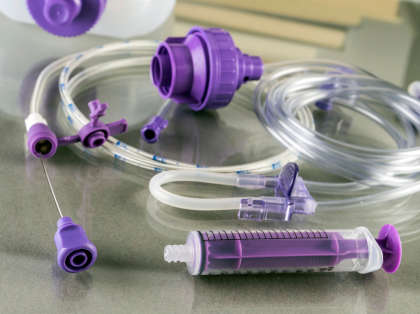
Living with TPN at home does not mean you are stuck at home. You can easily manage your nutrition independently or with the help of your caregiver. You can still lead a busy lifestyle. For instance, patients on total parenteral nutrition can still travel for business purposes as well as for social events and vacations. It just takes extra preparation, with guidance and support from your physician or medical team as well as your pharmacist. 

In fact, this transitional approach has significantly improved the quality of life of many patients

Familiar Environment

Patients who receive TPN at home benefit from being in comfortable, familiar surroundings. In addition to reducing stress, this can promote mental and emotional well-being. The total therapeutic experience might be enhanced by being close to loved ones.

Personalized Care and Cost-Effectiveness

For many patients, home-based TPN is more affordable than prolonged hospital stays and medical expenses. With home-based TPN, the financial burden that chronic illnesses frequently place on people and their families is lessened.

Healthcare professionals also adjust the TPN formulation according to the patient’s needs to ensure that patients receive optimal nutrition at home. This personalized care supports better overall health outcomes and increases the treatment effectiveness.

Challenges You May Face While Living With TPN at Home

Handling and Preparation of TPN Infusion

If you’re receiving TPN at home for the first time, it may be challenging to initially handle the technical aspects of TPN infusion. Generally, TPN-trained nurses educate the patient or their caregiver about handling, preparing, and administering the TPN mixture. However, patients still oftentimes find it challenging in the beginning. 

Sterility and Infection Risk

Another challenge you may experience while living with TPN at home is the increased risk of infection. Unlike in the hospital, where the TPN mixture is prepared and administered carefully in a sterile environment, receiving TPN at home may increase your risk of infections due to poor hygiene. 

Therefore, you need to follow strict hygiene practices during the preparation and administration of TPN to minimize the risk of potentially serious complications or catheter-related infections.

Speak to a Specialist About Copay Assistance

Schedule a Consultation

Tips To Manage TPN Infusion at Home

Here are some tips that you can follow to manage TPN infusions at home:

  • Ensure you or your caregiver gets comprehensive training and follow the provided instruction manuals about handling, preparing, and administering TPN mixture from your healthcare provider. 
  • When you start your TPN infusion cycle, ensure the TPN solution bag is in good condition, and there are no solid cloudy particles. 
  • Before preparing the solution, wash your hands with warm water and soap, and clean your space with alcohol wipes to avoid the risk of infection.
  • Prepare the TPN mixture according to the amount prescribed by your healthcare provider. Be sure to bring the TPN bag to room temperature before use to decrease the possibility of side effects.
  • Carefully insert the IV tubing into your catheter and IV infusion pump to avoid the risk of embolism.
  • Set your infusion at the prescribed rate and duration as instructed by your healthcare provider, and make sure to prime the IV tubing before starting. 
  • Typically, the TPN infusion takes 10 – 24 hours to complete. You can schedule your infusion cycle according to your daily routine, such as taking it in the morning or at night. 
  • Regularly monitor the infusion site and your central line before administering the TPN infusion.
  • Seek immediate medical attention if you see any severe redness or swelling at the site of injection or experience any side effects like diarrhea, fatigue, nausea, or headache. 
  • Keep in touch with your healthcare provider or attend follow-up appointments to discuss personalized guidance on managing TPN at home and assess your treatment’s effectiveness. 

Though home-based TPN comes with some challenges and requires adjustment to your daily activities and routine, with the right mindset, careful management skills, and healthcare guidance, you can enjoy your life while undergoing the treatment process at home. 

Ankylosing Spondylitis: Causes, Risks, Diagnosis, and Treatment

Ankylosing spondylitis (pronounced as ank-ee-lo-zing spon-dee-li-tus) or AS is a form of chronic inflammatory arthritis that predominantly affects your spine — a bony structure that provides your body shape and posture. The term “ankylosing” means stiffness; “spondyl” pertains to the spine or vertebra, and “itis” refers to inflammation. In essence, ankylosing spondylitis (AS) leads to inflammation in the spinal joints, which results in progressive stiffness and chronic pain, especially in the lower back. This inflammatory condition is also known as Bechterew’s disease.

Speak to a Specialist About Copay Assistance

(877) 778-0318

The symptoms of AS typically first show up in late adolescence or early adulthood. Men are three times more likely to get AS. In the United States, approximately 1.7 million adults have AS, according to the Centers for Disease Control and Prevention’s NHANES study. 

In this article, we will provide a brief overview of AS, including its symptoms, causes, risk factors, diagnosis, and treatment strategies for people with this condition or who are at a higher risk of developing it.

Before we delve into the basics of AS, it is important to understand this condition and how it affects your spine over time. 

Understanding Ankylosing Spondylitis

Your spine is a long column of bones that extend from your neck to your lower back. It is made up of several bones and soft tissues (i.e., collagen) that are joined together like bricks stacked on top of one another. Normally, your spine allows your body to move in every direction. For instance, the spinal joints attached to your hip bone, called sacroiliac joints, allow you to walk, twist and bend.

However, in the case of people with AS, the chronic inflammation of the joints in the spine makes the spine stiff. For example, daily activities like bending, walking, standing, and sitting become challenging for people with AS due to persistent lower back or joint pain. 

Moreover, in more advanced cases, inflammation can cause new bone formation (ankylosis) in which a section of the spine fuses together. This fusion leads to spinal deformities, and with time, the spine gradually loses its flexibility and mobility, which results in a hunched back and limited movement. 

Though it’s rare, AS also affects other large joints such as hips, shoulders, and, less often, ankles and knees. As the illness worsens, it may impact the joints connecting the ribs and spine, limiting chest movement and making it difficult to breathe deeply. 

What Are the Common Symptoms of Ankylosing Spondylitis?

Middle aged grey-haired woman suffering for back pain

The onset of AS symptoms can differ. For example, some individuals may experience mild episodes of pain, while others may experience chronic, severe pain. 

The most common early signs of AS are stiffness and frequent pain in the lower back and hip area (buttocks), which usually develops gradually over more than 3 months. Over time, the pain can spread up to the entire spine, into your neck, and may affect other parts of the body. 

Other symptoms include:

  • Stomach pain and diarrhea
  • Inflammatory bowel syndrome or bowel inflammation (seen in 50% of cases) 
  • Psoriasis (reported in 10% of cases)
  • Enthesitis
  • Dactylitis (inflammation in the entire finger or toe)
  • Anemia
  • Back pain and stiffness
  • Fatigue
  • Vision change and eye pain due to uveitis (seen in 25 – 30% of cases)
  • Weight loss and loss of appetite
  • Breathing difficulty if affected spinal joints connect to your ribs

Some people may feel more pain in the middle of the night or after extended sitting because the pain can worsen during rest or inactivity. Exercise and movement can typically help reduce discomfort. 
 

What Causes Ankylosing Spondylitis?

Though the exact cause of AS is unknown, researchers believe that both genetics and environmental factors play a role in developing this disease. Research shows that genetics increases your risk of developing AS. 

The human leukocyte antigen-B (HLA-B27) gene is a marker of AS, and 90% – 95% of people who develop AS have the HLA-B27 mutation gene. Your risk of AS increases if someone in your family or first-degree relatives has this condition.

However, in some rare cases, this gene is present but AS is never developed. This tells researchers that this is not a purely genetic disease. Environmental factors like a viral or bacterial infection may also need to be present to trigger the onset of AS disease in susceptible people. 

What Are the Risk Factors? 

Besides a genetic factor (human leukocyte antigen-B [HLA-B27] gene), there are other factors that may increase your risk of developing AS. These factors include:

  • Age: People younger than 40 are more likely to develop AS. For instance, around 80% of people experience the symptoms of AS before 30, and 5% are diagnosed with AS by the age of 40. 
  • Gender: AS is three times more common in males than females.
  • Race: Non-Hispanic white people are at higher risk of developing AS than other ethnic groups. 
  • Underlying diseases: People having other autoimmune conditions like Crohn’s disease, ulcerative colitis, and psoriasis are more likely to develop AS. 

How Ankylosing Spondylitis Is Diagnosed

Diagnosis of AS may be difficult since the presentation of back pain may be due to various health issues. Doctors may conduct multiple tests, as a single test cannot be used to diagnose accurately. In susceptible patients, rheumatologists do physical examinations and follow testing procedures. For example, a rheumatologist will ask:

  • About your symptoms, their severity, and how long you’ve had them
  • About your medical condition, like if you have an autoimmune disorder or any other disease
  • About your family history and if anyone in your family has AS

Based on your physical evaluation, if a rheumatologist suspects AS, then they will order genetic blood tests (for HLA-B27 gene detection) and take an imaging test, like an X-ray or MRI, to examine the appearance of your spine and pelvis, mainly the condition of the sacroiliac joint, which is the main joint that gets damaged in AS. 
 

Get Financial Assistance

Schedule a Consultation

How Is Ankylosing Spondylitis Treated?

Though there is no cure for AS yet, rheumatologists typically suggest particular treatment approaches that help in managing this condition and reducing the pain severity. 
Common treatments for AS include:

Medication

Certain medications like NSAIDs and biologic DMARDs are typically prescribed to ease pain and inflammation. This also helps to slow the progression of  AS symptoms.

Exercise

Since the symptoms of AS progressively improve with regular physical activity, exercise is typically recommended. Exercises like swimming, yoga, stretching, deep breathing, and posture practices can help maintain flexibility and range of motion. 

Surgery

Although rare, it is possible that in severe cases, a healthcare provider will recommend joint replacement surgery if a deformity is present in the knees or hip joints. People with poor posture due to fused bones may undergo an osteotomy. During this procedure, a surgeon will cut and realign the bones in the spine.
 

When To Visit a Doctor

If you have been experiencing severe back pain for the last 2 months or if your pain gets worse after you stop taking medications like NSAIDs, then you should consult your doctor immediately for an evaluation. 
The classical presentation of AS is that pain worsens at night or early in the morning (when you rest) and improves with exercise. 
 

Is There a Cure for Ankylosing Spondylitis?

Currently, there is no cure for ankylosing spondylitis. However, early diagnosis and effective treatment strategies such as medications, physical therapy, and good posture exercises can help reduce the symptoms and manage this condition. 
 

Conclusion

Ankylosing spondylitis (AS) is a rare form of chronic inflammatory arthritis that affects the spine as well as other joints throughout the body. The clinical presentation of AS is frequent pain and stiffness in the lower back and buttocks, which can worsen and affect other big joints if left untreated. This condition can be easily managed with early diagnosis and effective treatment strategies. 
 

What Is Ocular Myasthenia Gravis (OMG)?

Ocular myasthenia gravis (OMG) is a subtype of myasthenia gravis (MG) characterized by the weakening of specific muscles responsible for regulating the movement of your eyes and eyelids. Unlike myasthenia gravis, which broadly affects the skeletal muscles you can control, in ocular myasthenia gravis, only muscles surrounding your eyes and eyelids are affected. Eye muscle weakness can result in ptosis (drooping of eyelids), diplopia (double vision), and blurry vision.

Speak to a Specialist

About Copay Assistance
(877) 778-0318

In about 20 – 60% of cases, ocular myasthenia gravis could progress to generalized myasthenia gravis, where muscle weakness develops in other body areas such as the arms, legs, or throat. 

In this article, we’ll discuss the basics of OMG, how it occurs, its symptoms, diagnosis, and effective treatment options. 

Basics of Ocular Myasthenia Gravis

Ocular myasthenia gravis is an autoimmune condition in which your immune system produces auto-antibodies that target and destroy the connection between nerve and muscle cells. 

Generally, when a healthy person wants to move or blink their eyes, the brain sends nerve impulses or signals. The nerve endings then release a substance called acetylcholine that attaches to its receptor on the ocular (eye) muscle and causes a muscle contraction. 

However, in OMG, the auto-antibodies block the acetylcholine receptor sites and thus prevent signals from reaching the eye muscles to contract. Consequently, the eye muscles tire easily. 

What Are the Common Symptoms?

People with OMG experience trouble with eyesight due to drooping eyelids or double vision. The eyes do not move together in balanced alignment, so a single object may be seen as two images instead of one. Similarly, when the eyelids droop and cover some or all of the eye pupil, it obstructs the field of vision. 

Eye weakness can change from day to day, and these symptoms can worsen if the eyes are strained for a prolonged period of time. However, eye weakness can temporarily be improved if you rest and close your eyes when your symptoms are troubling you.

People with ocular myasthenia gravis DO NOT experience trouble breathing, speaking, or swallowing, nor do they experience weakness in the arms or legs. 

How Is Ocular Myasthenia Gravis Diagnosed?

Diagnosis involves a combination of clinical evaluation, specialized tests, and a review of the patient’s medical history. The following are some tests that doctors conduct to diagnose OMG:

  • Blood test to check for increased levels of autoantibodies
  • Electrical nerve testing to record the electrical signals sent with muscle movement
  • Repetitive nerve stimulation

Get IVIG Copay Assistance

IVIG Prior Authorization
Consult an IVIG Specialist

How Can You Treat Ocular Myasthenia Gravis?

The treatments can vary from person to person and may depend on the severity of symptoms. 

In most cases, doctors would prescribe a class of medication called cholinesterase inhibitors like Mestinon (pyridostigmine bromide). The medication helps to increase levels of acetylcholine to send signals to the muscle to move. If this medication is not effective then oral steroids like prednisone or other immunosuppressants are recommended to suppress the immune system. Other treatment options include:

Overall, the prognosis is good, and the symptoms are manageable. 

Conclusion

Ocular myasthenia gravis is an autoimmune condition that affects the function of the muscles that control the movement of your eyes and eyelids. This results in eye problems such as double vision or drooping eyelids. However, once diagnosed and treated, medication can help manage the condition and improve the quality of life.