Myasthenia Gravis Risk Factors: Who Is Most Likely Affected?

  • Myasthenia gravis is an autoimmune disorder causing muscle weakness due to impaired nerve-muscle communication.
  • Age and gender are key risk factors for myasthenia gravis, as it often affects younger women (<40) and older men (>60) more.
  • Other autoimmune diseases can increase the risk of developing or worsening MG.
  • Infections, certain medications, stress, and surgery are common triggers that can exacerbate myasthenia gravis symptoms.

Speak to a Specialist

About Copay Assistance
(877) 778-0318

Myasthenia gravis (MG) is a chronic autoimmune disorder characterized by weakness in the muscles responsible for movement. It is a relatively rare condition, with a prevalence of approximately 150 to 200 cases per million.

What are the risk factors for myasthenia gravis? Common myasthenia gravis risk factors include older age for men, younger age for women, and certain genetic markers.

In this guide, we will dive deeper into these factors. We will explain who is most at risk for myasthenia gravis and discuss the causes of this disease that you should be aware of.

What is Myasthenia Gravis?

Myasthenia gravis (MG) is a chronic autoimmune disorder that causes muscle weakness. This occurs when the communication between nerves and muscles is impaired.

Myasthenia gravis has several symptoms, some of which include:

  • Chronic fatigue
  • Difficulty walking
  • Drooping eyelids (ptosis)
  • Trouble holding up the head
  • Limited facial expressions
  • Double or blurred vision (diplopia)
  • Weakness in the neck, arms, or legs
  • Problems with swallowing or chewing
  • Difficulty speaking (soft or nasal speech)

Although there’s usually no cure, myasthenia gravis has several treatment options that can help improve symptoms.

What Causes Myasthenia Gravis?

The exact causes of myasthenia gravis are unknown. However, health experts believe specific genetic changes may make you more susceptible to developing this condition.

In myasthenia gravis, your immune system produces proteins (antibodies) that disrupt the communication between your nerves and muscles. Why this happens is not well understood.

Several studies have linked MG with thymoma (a rare tumor that forms on the thymus). That said, researchers do not yet know who is most at risk for myasthenia gravis and whether thymoma causes the disease.

What Are the Major Risk Factors for Myasthenia Gravis?

The major risk factors of myasthenia gravis include:

Age

Anyone can get MG, regardless of their age. However, women between 20 and 39 and men between 50 and 70 are more likely to develop this condition [1].

Gender

Like other autoimmune disorders, generalized myasthenia gravis affects more women than men. The female-to-male ratio of MG patients is about 2:1 [2].

Some women may experience their first symptoms during pregnancy. Many women report a worsening of symptoms before their menstrual cycle.

Thyroid

A person holding their wrist in pain

Having a thyroid condition is another risk factor for myasthenia gravis that may make you more likely to develop the disease. About 5% to 10% of people with MG have thyroid disease [3].

Thyroid conditions affect the thyroid gland in the neck, which produces hormones regulating metabolism. Hashimoto’s thyroiditis, which causes hypothyroidism (an underactive thyroid), and Graves’ disease, which results in hyperthyroidism (an overactive thyroid), are the most common conditions associated with MG.

Get Financial Assistance

Schedule a Consultation

Other Diseases

In rare cases, other autoimmune disorders may raise your risk of myasthenia gravis. Someone who is diagnosed with the following conditions may be at most risk for developing myasthenia gravis:

  • Rheumatoid arthritis (RA): Rheumatoid arthritis is a chronic inflammatory disorder that affects the joints, causing pain, swelling, and stiffness. However, it can also affect other parts, including the skin, eyes, lungs, heart, and blood vessels.
  • Systemic lupus erythematosus (SLE): Often called lupus, SLE is a chronic autoimmune disease that can affect various parts of the body, including the joints, skin, kidneys, blood cells, brain, heart, and lungs. It can cause widespread inflammation and tissue damage.

Myasthenia Gravis Risk Factors: Decoding the Role of Genetics

Genetic changes are thought to increase the risk of MG. However, these genes haven’t been identified yet.

Moreover, MG is not typically inherited and can occur in those with no family history. A family history of MG or other autoimmune conditions is present in only about 4% of the cases [4].

Rarely, children born to affected mothers get MG. This is known as neonatal myasthenia gravis. Neonatal MG is temporary. With appropriate treatment, complete recovery occurs within 60 days after birth.

Congenital myasthenic syndrome is a rare group of genetic conditions similar to myasthenia gravis but with different risk factors. It’s present in children at birth, with most symptoms beginning to show in childhood. However, in some cases, symptoms may not show until adulthood. It’s not an autoimmune condition, and its treatment differs from MG.

What Are the Other Potential Risk Factors for Myasthenia Gravis?

A 2023 study found that women over 50 who were diagnosed with type 2 diabetes may be at most risk for developing myasthenia gravis [5].

This research analyzed data from 118 hospitalized MG patients and compared them to several control groups, including the general population and patients with other autoimmune diseases.

The findings indicated that women over age 50 with type 2 diabetes had a significantly higher risk of developing MG. Interestingly, patients with diabetic MG tended to experience onset at an older age compared to those without diabetes.

The study suggests that diabetes may be one of the risk factors for myasthenia gravis, especially in older women. However, these are preliminary results. Further research is needed to confirm the link and understand the underlying mechanisms.

What Factors Can Make Myasthenia Gravis Worse?

If you have MG, certain factors can exacerbate your symptoms or trigger a worsening of your condition. Being aware of these triggers can help you manage your health better and avoid unnecessary complications. Here is an overview of these factors.

Infection

Infections can often worsen the symptoms of myasthenia gravis. When the body fights off infections, it naturally ramps up the immune response. This heightened response can intensify the autoimmune attack on the neuromuscular junction.

Common infections such as respiratory illnesses, urinary tract infections, or even minor colds can lead to increased weakness, fatigue, and in some cases, precipitate a myasthenic crisis.

Prompt treatment of infections and maintaining good hygiene are vital in minimizing this risk. If you notice your symptoms worsening during an infection, consult your healthcare provider immediately.

Get Copay Assistance Now

(877) 778-0318

Immunization

While vaccines are generally safe and essential for preventing serious diseases, certain immunizations can act as risk factors and temporarily exacerbate myasthenia gravis symptoms. Live vaccines or certain adjuvanted vaccines may trigger a flare-up.

The reason someone who is vaccinated is most likely at risk for experiencing a worsening of their myasthenia gravis symptoms is due to the immune system’s response to the vaccine, which can temporarily heighten autoimmune activity.

It’s important to discuss your condition with your healthcare provider before receiving any vaccination. They can advise on the safest options, timing, and whether premedication might be necessary.

In some cases, maintaining vaccination schedules is critical to prevent infections that could be risk factors for myasthenia gravis and worsen the patient’s condition.
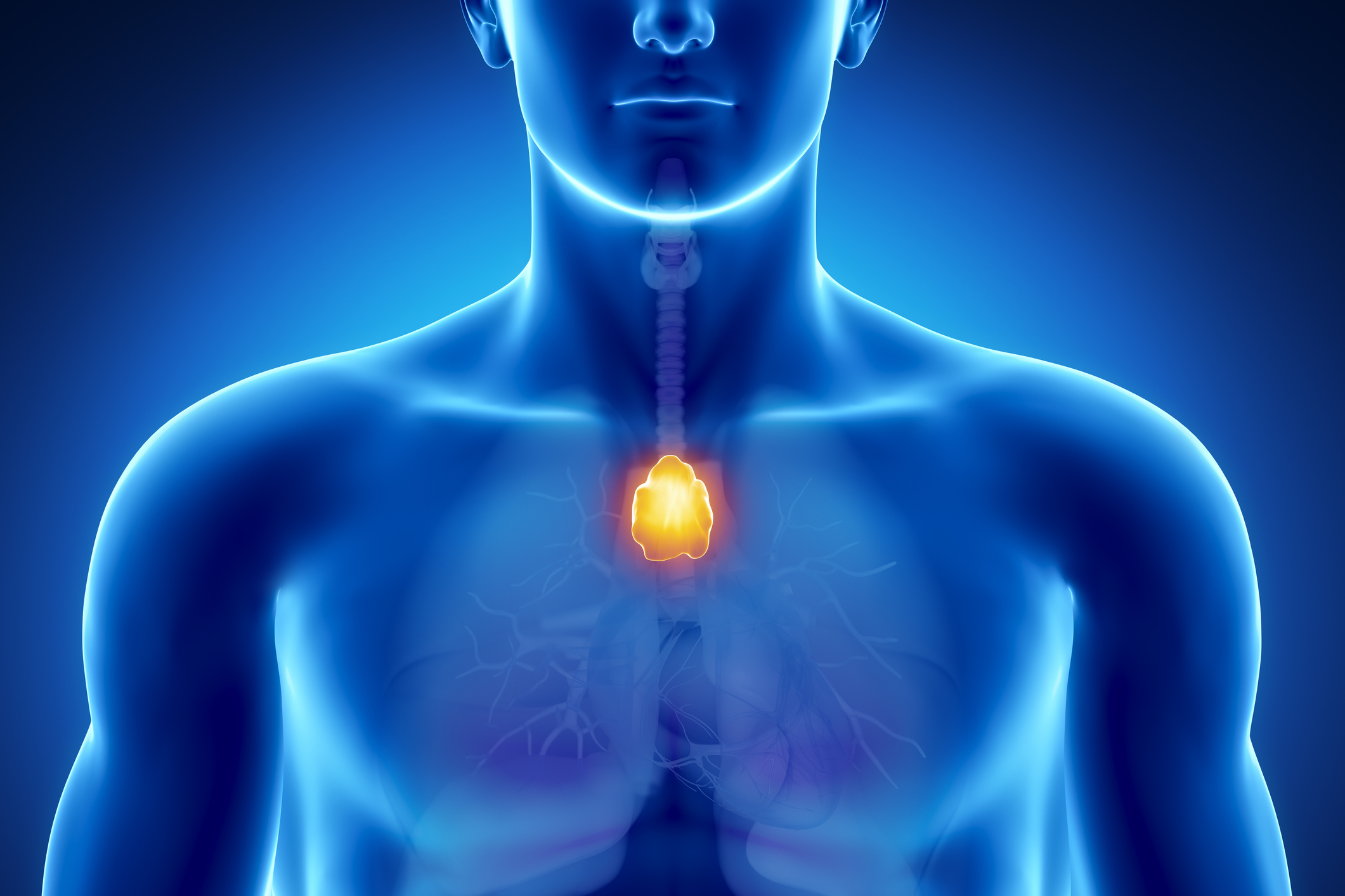
Surgery

Surgical procedures, especially those involving anesthesia or significant physiological stress, can impact MG symptoms. Surgery can cause physical stress and immune activation, temporarily worsening muscle weakness.

One treatment option for MG is thymectomy, which involves the removal of the thymus gland. However, this type of surgery requires careful planning before the operation and close monitoring afterward.

Anesthesia agents can also interfere with neuromuscular transmission. In these cases, anesthesiologists experienced with MG must be present during the operation.

If you’re scheduled to undergo surgery, it’s vital to inform your surgical team about your myasthenia gravis diagnosis so they can remedy factors that may risk exacerbating your condition. Proper perioperative management can minimize risks and ensure safer surgery.

Certain Drugs

Certain medications for other health conditions can interfere with neuromuscular transmission and worsen MG symptoms.

What are the drugs that can act as risk factors for exacerbating myasthenia gravis symptoms?

  • Certain antimalarial drugs (e.g., quinine and quinidine)
  • Seizure medications (e.g., phenytoin, carbamazepine, and phenobarbital)
  • Some antibiotics (e.g., aminoglycosides such as gentamicin and amikacin)
  • Medications for irregular heart rhythms (e.g., beta-blockers such as propranolol and metoprolol)
  • Statins for lowering cholesterol (e.g., atorvastatin, rosuvastatin, simvastatin)

It is best to avoid these drugs if you have MG. However, if needed, they should be used with caution and at the lowest dose possible. They can impair nerve-muscle communication, a factor that may increase the risk of myasthenia gravis symptoms worsening. Ask your doctor to recommend alternatives if possible.

Stress

Someone who is emotionally or physically stressed is also most likely at risk for exacerbating their myasthenia gravis symptoms.

Stress hormones can influence immune function and neuromuscular transmission, leading to increased weakness and fatigue. Prolonged or intense stress may also lower your threshold for experiencing a myasthenic crisis.

Developing stress management techniques such as relaxation exercises, mindfulness, and adequate rest can help reduce this risk. Maintaining a stable routine and seeking emotional support when needed are also helpful strategies to control your symptoms.

Myasthenia Gravis Risk Factors for Recurrence: What You Should Know

In some cases, the symptoms may reappear in people with MG who have had a thymectomy (surgery to remove the thymus). The risk is higher in [6]:

  • Males
  • Older adults
  • People with more severe disease

Death risk in MG patients may be higher if they have more severe symptoms, a stroke, diabetes, higher cholesterol levels, and abnormal heartbeats [7].

Speak to a Specialist

About Copay Assistance
Get Started

Myasthenic Crisis Risk Factors

A woman sitting on a couch holding her elbow in pain

Myasthenic crisis (MC) is a life-threatening complication of MG. It causes your breathing muscles to become too weak. You may need a breathing machine (ventilator) in severe cases.

MC is more likely to occur if you have [8]:

  • Thymoma
  • Higher antibody levels
  • Severe symptoms at diagnosis

Manage MG With AmeriPharma® Specialty Pharmacy

Understanding the risk factors for myasthenia gravis can help you better manage your condition and avoid complications. If you’re someone who is most at risk for developing myasthenia gravis, you might want to keep a close eye on emerging symptoms.

If you’re already diagnosed with MG and need treatment, AmeriPharma® Specialty Pharmacy can help. Our ACHC-accredited specialty pharmacy provides hard-to-find medications for those dealing with complex conditions like myasthenia gravis.

Contact us today to speak to a patient navigator and start receiving MG treatment with full-service coordination, copay assistance, and 24/7/365 support.

Toxic Epidermal Necrolysis (TEN): Causes, Symptoms, Treatment, and Survival Rate

Toxic epidermal necrolysis (TEN) is a rare, life-threatening skin reaction. It is most commonly triggered by certain medications. However, in rare cases, it can be caused by infections or vaccinations. 

Speak to a Specialist About Copay Assistance

Schedule a Consultation

A severe form of Steven-Johnson’s syndrome (SJS), TEN is diagnosed when you have large areas of blistering and peeling skin on more than 30% of your body. TEN also causes extensive damage to the moist linings (mucous membranes) in the mouth, nose, throat, eyes, and genitals.
In SJS, you have small areas of peeling skin, affecting less than 10% of your body. SJS-TEN overlap disease is when 10% to 30% of the body is affected.  

How Common Is Toxic Epidermal Necrolysis?

TEN is a rare condition, affecting 1.9 people per million U.S. adults each year [1]. 

What Causes Toxic Epidermal Necrolysis?

In some cases, the exact cause is not known. In others, several factors can induce TEN, including:

  • Antiseizure medications: Phenobarbital, phenytoin, carbamazepine, valproic acid, and lamotrigine
  • Antibacterial drugs: Sulfonamides (trimethoprim-sulfamethoxazole), chloramphenicol, penicillins, and ciprofloxacin
  • Non-steroidal anti-inflammatory drugs (NSAIDs): Piroxicam, meloxicam
  • Antigout agents: Allopurinol
  • Anti-HIV medications: Nevirapine and abacavir

Rare triggers can include:

  • Bacterial infections: Mycoplasma pneumoniae infections
  • Viral infections: Herpes and hepatitis A
  • Vaccines: Meningococcal B and MMR vaccines 
  • Graft-versus-host disease  

Risk Factors

The following factors can increase your risk of TEN:

  • HIV infection 
  • Weak immune system (such as in HIV/AIDS and autoimmune disorders)
  • Personal or family history of TEN
  • Genetic factors
  • Cancer (specifically blood cancers)

Symptoms

Mature woman suffering body aches from Toxic Epidermal NecrolysisSymptoms may appear a few weeks after you start taking a drug. Initially, you may have flu-like symptoms, such as:

  • Body aches
  • Cough 
  • Fever 
  • Chills
  • Headache
  • Red and swollen eyes

As the disease progresses, you may have:

  • Widespread skin pain
  • A rapidly spreading rash affecting more than 30% of your body
  • Blisters and peeling skin 
  • Sores and swelling on the mucous membranes, including the mouth, eyes, respiratory tract, and genitals

These signs and symptoms may resemble other skin diseases. Because TEN is life-threatening, seek immediate medical care if you think you have symptoms of TEN. 

Toxic Epidermal Necrolysis Treatment

People with TEN need emergency care at a hospital, preferably in an intensive care unit (ICU) or burn center. Treatment is mainly supportive and includes [2]:

  • Stopping all potential triggers
  • Isolation to prevent infection
  • Administering IV fluids and nutrition
  • Protective dressings and antibacterial creams or ointments to prevent skin infection
  • Antibiotic therapy if signs of infection are present
  • Medications to reduce pain
  • Eye and oral cavity examination 
  • Oxygen via a face mask (if needed)
  • Medications to prevent blood clots

No specific treatment for TEN exists. However, your provider may consider the following based on your age, symptom severity, expectations, and preferences:

Intravenous Immunoglobulin (IVIG)

IVIG is a sterile liquid that contains disease-fighting proteins (immunoglobulins or antibodies). Some studies show that early treatment with high-dose IVIG can improve survival and speed recovery [3].

Systemic steroids

Your provider may give you a steroid to halt the progression of TEN. Nonetheless, the use of steroids is highly controversial. Some studies suggest that steroids may increase the risk of death. 

IVIG With Steroids

According to a recent review published in the Journal of the American Academy of Dermatology, clinicians should consider combination therapy with IVIG and steroids to treat TEN [4]. The authors of the review conclude that this combination may reduce death risk in individuals with TEN. 

Plasmapheresis (Plasma Exchange)

Plasmapheresis separates the liquid part of the blood (plasma) from blood cells. Following separation, the blood cells are mixed with a liquid and infused back into the body. 
Daily plasmapheresis for 3 days can help remove the causative drug, its byproducts, and other inflammatory substances from the body. 

Tumor Necrosis Factor-Alpha (TNF-Alpha) Inhibitors

TNF-alpha inhibitors, such as infliximab and etanercept, may help resolve skin lesions in toxic epidermal necrolysis. They may be used alone or as a second-line medication.  

Can IVIG help? | Free IVIG Treatment Info

(877) 778-0318


Toxic Epidermal Necrolysis Survival Rate

Toxic epidermal necrolysis causes death in about 25% of the cases, and the death rate is lower in children (less than 10%). The death rate is higher if you [5, 6]:

  • Are over 40 
  • Have high blood sugar
  • Have a fast heart rate
  • Have chronic kidney disease


Complications

People older than 70 years are most likely to develop complications. Likewise, the risk of complications is higher in people with liver cirrhosis (scarring of the liver) or cancer. 
Complications of TEN include:

Sepsis

Sepsis or blood infection is a life-threatening condition. It happens when the immune response to an infection damages your body’s vital organs, putting you into shock and causing organ failure. 

Breathing Problems

If your lungs are involved, you may find it difficult to breathe. In severe cases, your lungs may be too weak, causing an excess of carbon dioxide and a deficiency of oxygen in your blood. 

Eye Problems

Eye problems associated with TEN are dry eye, ingrown eyelashes, corneal scarring, and, in rare cases, blindness.
 

Toxic Epidermal Necrolysis Long-Term Effects

Some long-term effects among TEN survivors are:

  • Dry itchy skin
  • Hair or nail loss
  • Excessive sweating
  • Dry eyes
  • Sensitivity to light 
  • Dry mouth
  • Lung damage

In a recent survey of 121 TEN/SJS survivors, more than half screened positive for depression, and over 40% screened positive for anxiety [7].
 

Get Financial Assistance

Schedule a Consultation


Toxic Epidermal Necrolysis (TEN) vs Steven-Johnson’s Syndrome (SJS)

The major differences between TEN and SJS are as follows:

Features

TEN

SJS

Severity

More severe

Less severe

Affected body surface

More than 30%

Less than 10%

Occurrence in the US

1.9 per million people each year

9.3 per million people each year

Death rate

About 25% in adults

About 10%


Frequently Asked Questions

Which drug causes toxic epidermal necrolysis?

Medications commonly associated with toxic epidermal necrolysis are:

  • Antiseizure medications: Phenobarbital, phenytoin, carbamazepine, valproic acid, and lamotrigine
  • Antibacterial drugs: Sulfonamides (trimethoprim-sulfamethoxazole), chloramphenicol,  penicillins, and ciprofloxacin

What causes epidermal necrolysis?

Medications are the primary causes of TEN in adults, whereas infections are the main causes in children.  

Which layer of skin is affected by toxic epidermal necrolysis?

Toxic epidermal necrolysis causes the top layer of your skin (epidermis) to detach from the lower layers of skin (dermis). 

What are the clinical features of toxic epidermal necrolysis?

TEN causes peeling skin on more than 30% of your body. It also causes extensive damage to the moist linings (mucous membranes) in the mouth, nose, throat, eyes, and genitals.
 

Hemlibra: A Promising Medication for Hemophilia A Patients

If you have hemophilia A, your doctor might suggest Hemlibra as a treatment option. Approved by the FDA on November 16, 2017, this medicine is used to prevent or reduce bleeding episodes in people with hemophilia A. Hemlibra is seen as a promising alternative to the usual treatments for this condition. In this article, we will discuss Hemlibra, its side effects, dosing information, and much more.

Speak to a Specialist About Copay Assistance

Schedule a Consultation

What Is Hemlibra?

Hemlibra (emicizumab-kxwh) is a medication made by Genentech, a subsidiary of the Roche corporation. This medication is used for preventing or reducing bleeding episodes in people with hemophilia A, whether they have factor VIII inhibitors or not. It was first approved by the FDA on November 16, 2017, for hemophilia A patients with factor VIII inhibitors [1]. Later, on October 4, 2018, the approval was extended for patients with hemophilia A without factor VIII inhibitors [2].

People with hemophilia A don’t have enough of a protein called factor VIII in their bodies.

This protein plays a very crucial role in the blood clotting process. Therefore, when someone with hemophilia A gets a cut or injury, their blood doesn’t clot properly. This can lead to longer bleeding, internal bleeding, and more problems.

Before the FDA approved Hemlibra, factor VIII replacement therapy was the primary treatment for hemophilia A. This therapy involved injecting external factor VIII proteins to compensate for the deficiency in hemophilia A patients. However, a common problem with this therapy is that, over time, some people develop inhibitors. Inhibitors are antibodies that attack factor VIII, making the treatment ineffective. Hemlibra offers a solution to this issue. It can prevent bleeding in people with hemophilia A, whether they have inhibitors or not. 

What Is Hemlibra Used To Treat?

Hemlibra is a breakthrough medication that is primarily used to treat hemophilia A. Hemophilia A is a rare genetic disorder that affects blood clotting due to a deficiency in clotting factor VIII. 

Hemlibra is prescribed to be taken regularly. It prevents or reduces bleeding episodes in patients of all ages, including newborns, with hemophilia A [3]. This medication is effective for both adults and children with this condition, whether they have factor VIII inhibitors or not. 

How Does Hemlibra Work?

Hemophilia A is a bleeding disorder caused by a missing clotting factor called factor VIII, which is a protein that helps in blood clotting. When you have an insufficient amount of factor VIII, your blood struggles to form clots when you have an injury. 

Hemlibra is an antibody that mimics factor VIII, the missing protein in hemophilia A.

Normally, factor VIII links factor IX and factor X to aid in blood clotting. However, in hemophilia A, this link is missing. Hemlibra fills this gap by bringing factor IX and factor X together, helping clot formation. This action reduces the danger of bleeding episodes, providing relief to people who have hemophilia A.

The usual treatment for hemophilia A is replacing factor VIII. However, some people may develop inhibitors over time, which can make this treatment ineffective. However, Hemlibra works uniquely. Rather than directly replacing factor VIII, Hemlibra performs the same function as factor VIII by joining other blood proteins. This means it remains effective, even if inhibitors are present. 

Side Effects

Like all medications, Hemlibra can also cause side effects. In rare cases, it can cause severe side effects. 

Common Side Effects

Woman with joint pain as a side effect of Hemlibra


The following are some common side effects:

  • Headache
  • Joint Pain
  • Reactions at the injection site, including redness, warmth, itching, or tenderness

These side effects usually go away on their own after a short time. But, if they worsen or do not go away, consult your doctor or pharmacist.

Severe Side Effects

Severe side effects from Hemlibra are rare, but they can occur. Contact your doctor right away if you encounter any severe side effects, which include:

  • Blood clots and injury to small blood vessels that cause harm to the kidney, brain, and other organs
  • Blood clots that may form in blood vessels in your arms, legs, lungs, or head
  • Stomach, chest, and back pain
  • Confusion and weakness
  • Swelling, pain, or redness
  • Eye pain or trouble seeing
  • Shortness of breath, coughing up blood

The above list of Hemlibra side effects is not exhaustive. If you encounter any other side effects that are not mentioned above, please consult your doctor. 

Get Financial Assistance

(877) 778-0318

Dosing Information

Hemlibra is administered subcutaneously through injections. The dosing depends on various factors like your weight and treatment history. Hemlibra treatment usually starts with loading doses, followed by maintenance doses. Loading doses rapidly increase the drug’s concentration in your body. They are higher than maintenance doses.

The first four doses are loading doses. They are given at 3 mg/kg of body weight once weekly. After that, each subsequent dose is known as a maintenance dose. They are given as:

  • 1.5 mg/kg of body weight once weekly, or
  • 3 mg/kg of body weight once every 2 weeks, or
  • 6 mg/kg of body weight once every 4 weeks

You have to discontinue the use of bypassing agents the day before starting Hemlibra treatment. If you miss a dose, take it as soon as you remember and then continue with your regular dosing schedule. 

Hemlibra Cost

Hemlibra can be expensive, priced at around $122 per mg [5]. The total cost of treatment varies based on your weight. For example, if you weigh 80 kg, the cost of treatment for the first year would be $411,136. For the following years, it would be $381,841. Please remember that this is a rough estimation. The real cost of treatment varies depending on your insurance plan, region, and pharmacy. 

FAQs

Here are some answers to frequently asked questions about Hemlibra:

Is Hemlibra Safe for Use During Pregnancy or Breastfeeding?

Hemlibra was mainly tested in males. There is no information about whether Hemlibra can cause harm to an unborn child or if it is present in breast milk. Hemlibra should only be used during pregnancy or breastfeeding if the benefits outweigh the risks.

Can Hemlibra Be Used in Patients With Liver or Kidney Disease?

If you have mild liver or kidney disease, you can still use Hemlibra without needing any changes to the dosage. Data for use in patients with severe liver or kidney disease is insufficient to provide specific recommendations [6]. Please consult your doctor.

Does Hemlibra Cure Hemophilia A?

Unfortunately, there is currently no cure for hemophilia A. Hemlibra can only prevent or reduce the occurrences of bleeding episodes. It cannot cure the disease. 

Hemophilia and Exercise: Tips for Staying Active and Healthy

Regular exercise benefits everyone, including those with hemophilia. However, for people with hemophilia, an exercise program needs to be customized based on:

  • Individual preferences and interests 
  • Age
  • Current activity level
  • Disease severity

Speak to a Specialist About Copay Assistance

(877) 778-0318


How Exercise Benefits People With Hemophilia

Regular physical activity improves joint function, reduces the risk of injury, and improves quality of life. Additionally, staying active can help improve pain, range of motion, and muscle strength.

Exercise strengthens bones and joints

Frequent joint bleeds are common among people with severe hemophilia. Over time, these types of bleeds can lead to joint swelling and destruction, which is associated with: 

  • Joint pain
  • Reduced mobility
  • Brittle bones
  • Reduced quality of life

Exercise helps preserve bone mass, reduces pain perception, and boosts joint movement [1,2].

Exercise strengthens muscles

Muscle bleeds can result in swelling, increased risk of infections, and loss of muscle strength [3].
Resistance training (weight lifting) not only increases muscle strength but also makes bleeding episodes less severe and less frequent [4].

Regular Exercise Helps Maintain a Healthy Weight

Obesity is a significant health issue among people with hemophilia. This is because hemophilic individuals tend to be more physically inactive. As a result, they are more likely to gain weight. 
According to the CDC Registry Report on Males with Hemophilia 2014-2017 [5]:

  • Among participants with hemophilia A aged 20 years or older, 34% were overweight, and 33% were obese.
  • Among participants with hemophilia B aged 20 years or older, 34% were overweight, and 37% were obese.

Both exercise and a healthy diet are effective tools for weight loss. Studies show that even moderate weight loss can decrease joint bleeds [6].
 

Hemophilia and Exercise: What Activities to Include and Avoid?

Water-based exercises may be more effective than land exercises for pain relief in adults [7].
For improving muscle strength, functional exercises seem to be more effective than static exercises. Examples of functional exercises are treadmill walking and partial weight-bearing exercises. 

Hemophilia activities list

Senior man with hemophilia in a swimming poolAccording to the National Hemophilia Foundation, safe activities include the following [8]:

  • Walking
  • Hiking
  • Swimming
  • Archery
  • Aquatics
  • Stationary bike
  • Elliptical machine
  • Fishing
  • Frisbee
  • Golf
  • Tai Chi
  • Snorkeling

Activities with safe to moderate risk include:

  • Bicycling
  • Using a rowing machine
  • Using a ski machine
  • Circuit training
  • Using the treadmill
  • Spinning
  • Physioball
  • Weight lifting


Get Financial Assistance

Schedule a Consultation

 
Activities with moderate risk include:

  • Aerobics
  • Bowling
  • Dance
  • Supervised recreational diving
  • Using a Stepper
  • Jumping rope
  • Roller-skating
  • Rowing
  • Tennis
  • Yoga

Activities with moderate-todangerous risk include:

  • Canoeing
  • Basketball 
  • Baseball
  • Gymnastics
  • Mountain biking
  • Kayaking
  • Karate
  • River rafting
  • Skateboarding
  • Soccer

You may choose to participate in these activities only after consulting your healthcare provider and with adequate precautions and supervision. 
Activities with highest risk (NOT RECOMMENDED) include:

  • Boxing
  • Competitive diving
  • Football
  • Hockey
  • Powerlifting
  • Lacrosse
  • Rock climbing in natural settings
  • Rodeo
  • Snowmobiling
  • Using the trampoline
  • Wrestling


Hemophilia and Exercise: How Long and How Often To Work Out?

The duration and frequency of exercise can vary depending on your fitness level, disease severity, type of activity, and goal. Regardless, any exercise program should routinely include the following steps: stretching, strengthening, conditioning, and aerobic training. In addition:

  • Warm up for about 10 minutes before starting your training. Cool down for 10 minutes after you finish your training. 
  • Perform strength training exercises two to three times a week with at least 48 hours rest between sessions. Each session typically lasts 30 minutes and should involve all major muscle groups. 
  • Do aerobic exercises for 15 to 20 minutes thrice weekly. Examples include walking, swimming, and riding a bike. If your target is weight loss, you should train for at least 30 minutes five times a week. 
  • Move as much as possible. Walk at a pace that allows you to speak but not sing for at least 30 minutes on most days of the week. 

Note: The recommendations above are for adults with hemophilia.
 

Hemophilia and Exercise: Tips for Optimal Results

  • Before starting a new activity, talk to your healthcare provider for an evaluation. This is critical even if you don’t have any specific health issues. 
  • Learn how to identify the early signs of a bleed (e.g., confusion, clammy skin, dizziness, light-headedness, weakness, shortness of breath). Seek immediate medical care if you notice one, especially if you suddenly feel any of these in relation to your exercise. 
  • You may receive prophylactic factor replacement before certain activities to help prevent bleeds. 
  • Choose activities you enjoy. Such activities are more likely to become a habit. 
  • Start with a low-impact and joint-friendly activity like swimming or stationary cycling. Once you gain enough strength, you may progress to harder ones. 
  • It is best to exercise with a friend or family member for added safety. 


Speak to a Specialist About Hemophilia

(877) 778-0318


What Sports Can You Play With Hemophilia?

Non-contact sports are safe for people with hemophilia. Examples include swimming, walking, and jogging. Participation in demanding sports such as rugby, boxing, football, and basketball is not recommended. 
 

A Comprehensive Guide to Autoimmune Encephalitis

In the world of brain health, there’s a lesser-known condition called autoimmune encephalitis. In this condition, your body’s immune system mistakenly attacks your brain cells. This causes inflammation and a wide range of neurological issues, from memory loss to mood swings. If left untreated, this condition can even lead to death. But, with early diagnosis and treatment, you can recover. In this article, we will discuss autoimmune encephalitis, its symptoms, diagnosis, treatment, and much more.

Get Your IVIG Dose – At-Home Infusion

(877) 778-0318

What Is Autoimmune Encephalitis?

Autoimmune encephalitis is a group of disorders in which the body’s immune system mistakenly attacks healthy brain cells. This causes inflammation in the brain which disrupts the brain’s normal function and leads to a wide range of neurological symptoms. 

In the past, this condition was thought to be rare, but now it is being diagnosed more frequently thanks to improvements in medical techniques. A recent study found that approximately 13.7 cases occur per 100,000 people [1]. Also, this disease is more common in females than in men. 

Without treatment, this condition can quickly become serious. It can possibly lead to coma or permanent brain damage. In rare cases, it can cause death. So, early diagnosis and treatment are crucial for proper recovery. 

Symptoms of Autoimmune Encephalitis

Autoimmune encephalitis can cause a variety of signs and symptoms. Early symptoms of this disease appear within a few weeks to 3 months [3]. They include symptoms like headache, fever, and memory loss. The later symptoms can be more severe. They include symptoms like impaired consciousness, hallucinations, and personality changes.

Here are some other common symptoms:

  • Memory problems, confusion, difficulty concentrating
  • Hallucinations, delusions, paranoia, mood swings, anxiety
  • Seizures
  • Abnormal and involuntary movement
  • Speech and language difficulties, such as difficulty finding words or trouble understanding language
  • Drowsiness 
  • Insomnia
  • Weakness or numbness
  • Headache

Please consult your doctor if you are experiencing any symptoms mentioned above for proper diagnosis and management. 

What Causes Autoimmune Encephalitis?

Autoimmune encephalitis is a complex disorder. What exactly causes this condition is not always clear. A wide range of factors can cause this condition:

  • In many cases, for unknown reasons, your immune system can produce antibodies that target your brain cells. This can lead to autoimmune encephalitis.
  • Certain bacteria and viruses like HSV (herpes simplex virus) or streptococcus can trigger an immune response that mistakenly targets brain cells [3]. This can also lead to autoimmune encephalitis.
  • Certain tumors like teratoma (ovarian tumor) can trigger an immune response that targets brain cells.
  • Rarely, there may be genetic factors that contribute to the development of this condition [4].
  • Certain environmental factors, like exposure to certain toxins or chemicals, may play a role in triggering autoimmune responses [5]. 

Can IVIG help? | Free IVIG Treatment Info

Schedule a Consultation

Diagnosis

Brain MRI scan

Autoimmune encephalitis is difficult to diagnose, and often it is misdiagnosed. Research suggests that up to 72% of cases may be initially misdiagnosed [6]. The reason for this is that doctors consider autoimmune encephalitis as rare, and they mistake it for some other illness. 

Early diagnosis is very crucial for this condition. If left untreated for a long time, it can be fatal. The diagnosis includes the following:
Clinical Evaluation: Doctors will examine your medical history and symptoms. If they suspect you have this condition, they will conduct further medical tests.
Blood Test: Doctors conduct blood tests to see if your blood contains any inflammatory markers or antibodies associated with autoimmune encephalitis.
Lumbar Puncture: A lumbar puncture or spinal tap is performed to withdraw a sample of your cerebrospinal fluid. This sample is then analyzed for signs of inflammation and antibodies.
MRI Scan: Doctors perform MRI scans of your brain to look for signs that indicate this condition.
Autoantibody Testing: Some specialized tests are performed to detect the presence of autoantibodies associated with autoimmune encephalitis. 

Treatment

For autoimmune encephalitis, early treatment is crucial. Early treatment can make a big difference in reducing your symptoms and preventing long-term complications. The treatment you’ll receive will depend on your specific symptoms, disease severity, underlying causes, and response to therapy. In general, the treatment of autoimmune encephalitis includes:

Steroids: These are anti-inflammatory medications that can reduce the immune system’s response and brain inflammation.
Intravenous Immunoglobulin (IVIG): IVIG contains antibodies obtained from the plasma of healthy donors. It works by destroying harmful antibodies, which in turn helps to decrease inflammation in the body.
Plasma Exchange: In this procedure, your blood plasma is replaced with a healthy donor’s plasma. This helps to get rid of harmful antibodies.
Immunosuppressive Medications: These medicines are used to suppress the immune system’s activity and prevent further attacks on the brain.
Surgery: If a tumor causes your condition, then your doctor will perform surgery to remove it.
Pain and Seizure Management: Doctors will recommend medications to control seizures and manage your pain.
Physical Therapy: Doctors may recommend physical therapy. It can help improve your motor skills, communication, and overall functioning. 

Get IVIG Copay Assistance – Speak to a Specialist

(877) 778-0318

FAQs

1. Can Autoimmune Encephalitis Be Cured?

In some cases, it can be cured with proper treatment. The recovery depends on several factors, including one’s underlying cause, severity of the symptoms, and response to treatment. Some people recover quickly within months of starting treatment. For others, recovery may take years [7].

2. Can Autoimmune Encephalitis Recur?

Yes. Autoimmune encephalitis can recur after successful treatment. So, close monitoring and follow-up care are important to detect any recurrence early [8].

3. Are There Any Long-Term Effects?

Some people may experience long-term effects even after treatment. Various symptoms may last for many years. These symptoms include seizures, problems with memory and judgment, fatigue, sleep disturbances, depression, and personality changes [9]. You may need ongoing medical care to manage these symptoms and improve your quality of life.

4. Is There Anything I Can Do to Reduce My Risk?

The exact cause of autoimmune encephalitis is not always clear. Still, there are some steps you can follow to reduce your risk. You should maintain overall good health, avoid known triggers such as certain infections or toxins, and seek immediate medical help if you notice any neurological symptoms. 

Exploring Hemophilia Risk Factors: From Genetic Inheritance to Acquired Triggers

Knowing about hemophilia risk factors can help reduce your risk of developing complications.

Speak to a Specialist About Copay Assistance

Schedule a Consultation

What Is Hemophilia?

Hemophilia is a group of hereditary bleeding disorders. It happens when your blood contains little or no blood-clotting proteins called clotting factors. As a result, your blood cannot clot properly, which leads to excessive bleeding. 

The exact number of Americans living with hemophilia has yet to be determined. The Centers for Disease Control and Prevention (CDC) estimates that 33,000 males in the United States suffer from this disorder. Nearly 400 babies are diagnosed with hemophilia A (a type of hemophilia) at birth each year [1].

What Are the Complications of Hemophilia?

  • Excessive and prolonged bleeding, even without an injury or trauma
  • Internal bleeding (bleeding in a vital organ such as the brain can be fatal)
  • Bleeding into the joints (can cause chronic joint pain and swelling)

Hemophilia Types

Several different types of hemophilia are described in medical literature. Among them, the two most common are:

  • Hemophilia A (Classic Hemophilia): People with hemophilia A have little or no clotting factor VIII in their blood. 
  • Hemophilia B (Christmas Disease): People with hemophilia B have little or no clotting factor IX in their blood.

A rare form, hemophilia C, can occur due to a lack of clotting factor XI. 

Hemophilia Risk Factors: 3 Things You Should Know

1. Genetics Plays a Crucial Role in All Three Types of Hemophilia

In over 66% of all hemophilia cases, there is a family history of the condition [1]. Thus, having one or more family members with hemophilia is a major risk factor. 

Hemophilia occurs when you have a faulty gene on your X chromosome. 

Men with the faulty gene (and hemophilia) pass it on to their daughters. Also called the “carriers,” the daughters may or may not develop hemophilia. However, the sons don’t inherit the faulty genes. 

If women carry faulty genes, there is a 50% chance their [2]:

  • Sons will have hemophilia
  • Daughters will be a carrier

2. Most People With Hemophilia Are Males

Men have only one X chromosome. So, any defective gene on the X chromosome means they will have hemophilia. Understandably, the majority of patients with hemophilia are male.

On the other hand, women have two copies of the X chromosome. So, even if they have a genetic defect on one chromosome, their bodies can make enough clotting factors with instructions from the healthy gene on the other chromosome. 

3. In About One-Third of Cases, There Is No Family History

Hemophilia patient with bandages on knee

Hemophilia is almost always inherited. However, some people may develop this disorder even though they have no family history of the condition. This type is known as acquired hemophilia. 

It is a rare disorder that affects nearly 1.5 in every million people [3]. The risk is similar in both males and females. No causes can be identified in about 50% of cases. In others, the acquired hemophilia risk factors include: 

Get Financial Assistance

(877) 778-0318

What Are the Risk Factors for von Willebrand disease?

People with von Willebrand disease (VWD) lack a blood-clotting protein called von Willebrand factor (VWF). It is a common inherited blood disorder that affects 1% of Americans [4].

Genetics, which means having someone in your family with the disorder, is the most common risk factor. 

What Are the Risk Factors for Bleeding Disorders?

The following factors may make you more likely to get bleeding disorders:

Age

Bleeding disorders can affect people of any age. However, newborns have a higher risk of developing vitamin K deficiency bleeding. Likewise, older adults are at a higher risk of acquired hemophilia. 

Family history

You are more likely to have a bleeding disorder if one or both of your parents have the disorder. 

Sex

Hemophilia is far more common in males. On the other hand, the risk appears similar for men and women for certain bleeding disorders, such as acquired hemophilia.

Clinimix vs. TPN: How Are They Different?

Clinimix is a preformulated total parenteral nutrition (TPN) intravenous formula. It provides nutritional support to patients who are unable to obtain adequate nutrients orally due to gastrointestinal dysfunction or other illnesses. The purpose of TPN is to fulfill the nutritional requirements of patients. TPNs can differ in their composition and storage requirements. This article discusses the key differences between Clinimix and a customized TPN and when each type of formula is suitable for patients.

Always in Stock

Full inventory of IV fluids, ready for you
(877) 778-0318

Clinimix vs. TPN: A Basic Understanding

What Is Clinimix?

Clinimix is a brand name for a premixed total parenteral nutrition solution manufactured by Baxter Healthcare. It is a ready-to-use nutrition solution formulated with up to 80 grams of amino acids per liter. Clinimix specifically provides macronutrients (amino acids and dextrose) and micronutrients (electrolytes) by intravenous infusion. 

It is a nutritional premix containing essential ingredients that can be used as an alternative to customized TPN. This makes Clinimix a convenient option for healthcare providers to choose Clinimix formulations for patients who require moderate to high protein content. 

What Is TPN?

Unlike Clinimix, TPN is a customized formula that can provide more nutrients such as carbohydrates, proteins, fats, vitamins, and minerals to patients intravenously (through veins) in a balanced ratio. The formulation of total parenteral solution is determined by healthcare providers or pharmacists according to the patient’s complex nutritional requirements.

Clinimix vs. Custom TPN: Key Differences

Clinimix and custom TPN differ in various aspects, such as indication (usage), composition, storage, and contraindication. 

Indication

Clinimix is given when patients require moderate to high sources of calories and protein. Their diet does not provide enough calories and protein, usually due to certain illnesses or recent surgery. Clinimix comes in various formulations and can contain up to 80 g/l of amino acids. It is also used in critically ill adult patients with significant lean body mass in the first 7 – 10 days of ICU stay. Clinimix aims to reach patient protein targets to meet their nutritional goals.

Moreover, Clinimix treats patients with negative nitrogen balance in the blood. 

Unlike Clinimix, TPN is used for patients who require complete nutritional support when oral or enteral feeding is not feasible or contraindicated. It is often indicated for patients with severe malnutrition, gastrointestinal disorders, or those undergoing major surgeries.

Composition & Formulation

The composition and formulation of Clinimix are different from that of TPN. Clinimix is available in fixed formulations and concentrations and cannot be customized. Currently, Clinimix is available in two forms: 

  • Clinimix: This formulation contains macronutrients like proteins in the form of essential and non-essential amino acids and carbohydrates (dextrose).
  • Clinimix E: This formulation contains essential and non-essential amino acids with electrolytes and dextrose with calcium. 

Clinimix is available in 1 and 2 liters of volume with fixed formulations of 8/14 (8% amino acids and 14% dextrose), 8/10 (8% amino acids and 10% dextrose), and 6/5 (6% amino acids and 5% dextrose) for central and peripheral infusions. 

In contrast, TPN is a custom-prepared solution that contains a precise balance of carbohydrates, proteins, fats, vitamins, and minerals tailored to meet the specific nutritional requirements of individual patients.

Storage

Clinimix is stored at room temperature (25°C/77°F), while TPN is stored in the refrigerator at a temperature of 2 – 8°C. 

Availability

Since TPN is a custom-prepared solution, it is typically prepared by pharmacies within hospitals or specialized compounding facilities.

On the other hand, Clinimix solutions bags are commercially available to be dispensed by pharmacies. 

Speak to a Specialist About Copay Assistance

Schedule a Consultation

Contraindications

TPN is contraindicated under the following cases:

  • Patients with cardiovascular instability or metabolic instabilities
  • When gastrointestinal feeding is possible
  • Infants with less than 8 cm of the small bowel
  • Patients with good nutritional status 

In contrast, Clinimix and Clinimix E are contraindicated in the following cases:

  • When patients have amino acid metabolism concerns
  • Patients with pulmonary edema or acidosis due to low cardiac output
  • Patients with known hypersensitivity to one or more amino acids or dextrose
  • Neonates (28 days of age or younger) receiving concomitant treatment with ceftriaxone

In conclusion, the purpose of Clinimix and TPN is to provide nutritional support to patients. However, they differ in terms of composition, formulation, usage, storage, and contraindications. For example, a custom TPN provides complete nutritional needs. It is prepared manually based on the patient’s requirements. Conversely, Clinimix is a ready-to-use nutritional solution with higher protein and less dextrose, intended for patients with moderate to high protein requirements. The selection of each method depends on the patient’s clinical condition and nutritional requirements. 

Understanding Birdshot Retinochoroidopathy: Symptoms, Diagnosis, and Treatment Options

Birdshot retinochoroidopathy, also known as birdshot uveitis, is a chronic disease of the eye. This condition is rare and primarily affects the two crucial components of your eye, the retina and choroid, which are responsible for vision. 

Speak to a Specialist About Copay Assistance

(877) 778-0318

Birdshot retinochoroidopathy can occur at any age but is predominantly seen in middle-aged people. Research shows that females are more prone to developing this eye condition than males. 
Though this disease is uncommon, understanding this condition’s onset can help you manage its symptoms through immediate treatment. 
In this article, we will briefly explain what birdshot retinochoroidopathy is, how it occurs, its symptoms, prevalence, diagnosis, and available treatment options.

Overview of Birdshot Retinochoroidopathy

This inflammatory eye disease was initially identified in 1949. The condition was later called “birdshot retinochoroidopathy” by two eye specialists, Ryan and Maumenee, in 1980 because of its unusual clinical appearance, which consisted of cream-colored oval spots in the superficial (top layer) choroid and deeper retinal layer observed in 15 patients. These dots are similar in pattern to small shotgun pellets or birdshots (bullets).
Birdshot retinochoroidopathy is also known by other terms, such as birdshot chorioretinopathy and vitiliginous chorioretinitis, to name a few.  

How Birdshot Retinochoroidopathy Occurs

The exact pathogenesis of birdshot retinochoroidopathy is unknown. However, it is suspected that it may occur as a result of an immune attack, where your immune system mistakenly attacks your healthy tissues (in this case, the cells of your retina and choroid). 
The retina is the light-sensitive tissue at the back of your eye that senses light signals and sends signals to the brain so you can see. Choroid, which is packed with blood vessels to provide eyes with oxygen and nutrients, connects your retina to the white part of your eye. 
The immune attack causes inflammation in these areas (retina and choroid), which leads to visual disturbance and vision loss over time. 
Genetic factors may also play a role in the onset of this condition. According to research, the HLA-A29 gene variant triggers immune reactions and is central in the development of birdshot retinochoroidopathy. Around 95% of birdshot retinochoroidopathy patients carry the HLA-A29 allele. 
However, HLA-A29 positivity doesn’t mean you’ll definitely develop the condition since birdshot retinochoroidopathy is also reported in non-HLA-A29 carriers. Therefore, possessing the HLA-A29 allele alone is not sufficient for a diagnosis. 

What Are the Symptoms of Birdshot Retinochoroidopathy?

An ophthalmologist examines a woman's eyes
The symptoms of birdshot retinochoroidopathy may vary among patients. Since it is a chronic (long-term) condition, symptoms develop and progressively get worse over time (several months to years). 
Birdshot retinochoroidopathy mainly affects both eyes. The early symptoms you are most likely to experience are blurred vision, floaters (seen in 29% of cases), and decreased vision (seen in 68% of cases). 
However, you may also experience the following symptoms:

  • High eye pressure (ocular hypertension)
  • Night blindness (nyctalopia, seen in 25% of cases)
  • Problems with color vision (dyschromatopsia, seen in 20% of cases)
  • Sensitivity to bright lights (photophobia)
  • Seeing flashing lights
  • Distortions in shapes
  • Loss of depth perception or peripheral (side) vision

In severe cases, you may experience cataracts, which can cause severe vision loss even after treating inflammation. It’s essential to consult your eye specialist if you experience the symptoms mentioned above. 

How Common Is Birdshot Retinochoroidopathy?

Birdshot retinochoroidopathy is a rare disease and accounts for 1 – 2% of all types of uveitis. It is more prevalent in middle age (between 40 and 60 years of age) and rarely affects children. 
The incidence of this disease is highly prevalent in White populations. 
Gender-wise, women are more likely to develop this condition than men. 

Get Financial Assistance

Schedule a Consultation


How Is Birdshot Retinochoroidopathy Diagnosed?

The diagnosis of birdshot retinochoroidopathy can be difficult. It is a rare disease, and unique spots may not be visible in the early stages. 
However, based on the symptoms of the patients, eye specialists examine the eyes, and if they suspect you have this condition, they may run some clinical and functional testing, which includes:

  • Fluorescein angiography (to check retinal inflammation)
  • Fundus autofluorescence (to check photoreceptor health)
  • Optical coherence tomography (to analyze the retinal and choroidal layers)
  • Blood test (to check HLA-A29 antigen in the patient’s blood) 

These tests are also commonly used to monitor the progression of the disease over time. 
 

What Are the Treatment Options for Birdshot Retinochoroidopathy?

Currently, there is no available cure. Initially, Eye specialists prescribe high doses of corticosteroids to control eye inflammation and then slowly reduce the dose due to their side effects. 
These corticosteroids could be administered as eye drops, injections, or oral medicines to reduce the inflammation. 
In some cases, you may also require long-term treatment with immunosuppressant drugs or immunomodulatory therapy (IMT), a standard care treatment for birdshot retinochoroidopathy. This therapy stops your immune system from attacking your eyes, prevents inflammation, and preserves visual function. 
 

Conclusion

Birdshot retinochoroidopathy is an eye disease that causes inflammation in the retina and choroid. Patients experience blurred vision, floaters, or decreased vision. It mostly affects middle-aged people and is more prevalent in females. This condition can even lead to permanent vision loss if undertreated or unrecognized. 
Early on-time diagnosis and effective treatment regimes can help slow the disease’s progression and manage its symptoms. 
 

Myasthenia Gravis and Thymoma: Exploring the Link and Treatment Options

Myasthenia gravis (MG) is an autoimmune disease that causes muscle weakness. In this condition, your body’s defense system mistakenly attacks nerve receptors. As a result, your eyelids, eye movements, breathing, swallowing, and limbs are affected. 

Speak to a Specialist About Copay Assistance

(877) 778-0318

Thymoma is a rare tumor that grows in a gland called the thymus. There is a link between myasthenia gravis and thymoma. Many people with MG also have thymoma and vice versa. In this article, we will learn about myasthenia gravis and thymoma, their connection, and treatment options. 

What Is Myasthenia Gravis (MG)?

Myasthenia gravis (MG) is a rare progressive autoimmune disease. Every year, it affects up to 30 persons in every 1 million [1]. In this condition, the body’s immune system mistakenly attacks the receptors on muscle cells. As a result, the signals from the brain can’t reach the muscles effectively. This leads to weakness and fatigue in the facial, cranial, and bodily muscles.  

The symptoms of MG can vary in severity. People experience increasing muscle weakness during activity, which is relieved during rest. There is no cure, but there are options like medication, therapy, and surgery to manage the symptoms and improve the quality of life.

What Is Thymoma?

Thymoma is a rare type of tumor that develops in the thymus gland. The thymus gland is located in the chest, just behind the breastbone. This gland plays a crucial role in the body’s immune system. It promotes the maturation of T cells, which are a type of white blood cell that fights infections. 

Thymomas usually grow slowly, and it can take months or years to see significant growth. They do not cause any symptoms in the early stages. However, as they grow large, they can cause symptoms like chest pain, coughing, difficulty breathing, etc. 

What Is the Link Between Myasthenia Gravis and Thymoma?

Myasthenia Gravis (MG) is a disease in which the body’s immune system attacks areas where the nerves communicate with muscles. As a result, the lack of communication causes muscle weakness and fatigue. On the other hand, a thymoma is a rare tumor that grows in the thymus gland. This gland helps the immune system to work properly.

Thymoma is often associated with myasthenia gravis (MG). Data shows that about half of the thymoma patients develop MG, and about 15% of MG patients develop thymoma [2].

However, doctors do not exactly know whether thymoma causes MG or MG causes thymoma. However, a compelling theory suggests that when there is a problem with the thymus gland (like thymoma), it sends confusing signals to the immune system. As a result, the immune system attacks the receptors on the muscle cells, and thus MG occurs. 

Thymomas are more common in patients who develop MG at a younger age, between 30 and 60. If MG starts when someone is older than 60, they are less likely to have a thymoma [3]. 

Symptoms of Myasthenia Gravis and Thymoma

Woman with chest pain from thymoma

The symptoms of myasthenia gravis (MG) and thymoma can vary depending on the individual and the severity of the condition.

MG causes muscle weakness that gets worse with activity and improves with rest. This leads to symptoms like:

  • Trouble eating or swallowing
  • Blurred vision
  • Drooping eyelid
  • Arm and leg weakness
  • Slurred speech
  • Trouble breathing

People with MG must undergo tests like X-rays to discover thymoma’s presence. In the initial stages, thymoma doesn’t show any symptoms. As its size gets bigger over time, it may lead to symptoms like:

  • Chest pain
  • Coughing
  • Difficulty breathing 
  • Difficulty swallowing 

Please consult a healthcare professional if you have symptoms related to either condition. They can give you a proper diagnosis and treatment. 

Get IVIG Copay Assistance | IVIG Financial Assistance

Consult an IVIG Specialist

Treatment of Myasthenia Gravis and Thymoma

For myasthenia gravis, there is no cure. However, some treatment options exist to manage the symptoms and enhance quality of life. Treatment options for MG include:

Medications: Medications can improve muscle strength and reduce symptoms. Doctors commonly prescribe medicines like cholinesterase inhibitors, corticosteroids, and monoclonal antibodies. 

Plasma Exchange: This is a procedure to remove harmful antibodies from your blood plasma.

Intravenous Immunoglobulin (IVIG): IVIG provides a concentrated dose of antibodies obtained from donated blood plasma. It can help reduce the symptoms of MG and improve muscle strength.

Surgery: Whether or not you have a thymoma, surgical removal of the thymus gland can help a lot in cases of MG [5].

Treatment for thymoma typically involves surgery. If the thymoma is non-cancerous, then surgical removal of the thymus gland is enough. However, if the thymoma is cancerous, additional treatment, such as chemotherapy, is recommended along with surgery. 

FAQs

Here are some frequently asked questions about myasthenia gravis and thymoma:

1. How Is Myasthenia Gravis Diagnosed?

To diagnose myasthenia gravis, doctors use a combination of tests. Diagnosis often involves blood tests to check for specific antibodies. It also involves nerve conduction studies and electromyography (EMG).

2. Is Thymoma Always Cancerous?

No. Thymomas can be benign (non-cancerous) or malignant (cancerous). Fortunately, the majority of them are non-cancerous. In both cases, you need regular monitoring and proper treatment.

3. Are There Any Lifestyle Changes That Can Help Manage Myasthenia Gravis Symptoms?

Yes, MG patients can live better lives if they learn to manage their stress. They should get adequate sleep, eat a balanced diet, and avoid things that worsen their symptoms (such as certain medications or extreme temperatures).

4. Does Myasthenia Gravis Affect Pregnancy?

Myasthenia gravis (MG) typically does not cause significant problems during pregnancy. Reports suggest that women with MG do not have a higher chance of miscarriage or premature delivery [4]. 

CIDP and Physical Therapy: How Supervised Exercises Can Help Improve Quality of Life

Chronic inflammatory demyelinating polyneuropathy (CIDP) reduces your ability to perform activities that are considered “standard” or “normal” for a healthy person to complete. This is called exercise intolerance. Physical therapy uses individualized exercise programs to help you carry out daily activities. 

Speak to a Specialist About Copay Assistance

(877) 778-0318

CIDP and Physical Therapy: Understanding the Basics

CIDP (chronic inflammatory demyelinating polyneuropathy) is a neurological disorder. 

People with this condition experience progressive loss of muscle strength and senses in their arms and legs. As a result, they have problems with balance and gait, which can affect their ability to perform their normal daily activities. 

CIDP also raises your chances of developing depression and anxiety [1]. If you have symptoms of these conditions, seek immediate medical care. 

Medical treatments for CIDP include immunosuppressants, steroids, IVIG, and plasmapheresis. Rehabilitative interventions include physical therapy, occupational therapy, adaptive equipment, and emotional support. Medical and rehabilitative therapies go hand-in-hand and are typically used together. 

Physical therapy is a critical component of rehabilitative intervention that can help:

  • Maintain muscle strength 
  • Prevent complications such as contractures or respiratory failure
  • Improve mobility and functional independence

Physical therapy uses specific exercises and physical activities to help you move better and improve muscle strength. 
 

CIDP and Physical Therapy: What To Expect

Before recommending a CIDP exercise program, your physical therapist will assess your:

  • Strength
  • Baseline fitness level
  • Perceived barriers to exercise

Based on this information, they will customize exercises for CIDP to help you meet your unique needs. 
 

CIDP and Physical Therapy: What Are the Benefits?

Physical therapy benefits anyone with CIDP, regardless of the stage (severity) of the disease. 

Physical Therapy During a Flare-up

Nurse helping CIDP patient in wheelchair

Most people are unable to move during a CIDP flare-up. In such cases, your physical therapist will educate you and your caregiver about ways to prevent muscle tightness and bedsores. These include:

  • Avoiding prolonged hip and knee flexion (bending)
  • Changing your position every two hours in bed
  • Supporting weak upper limbs using armrests or pillows

In addition, they will teach you how to use assisted devices and perform breathing exercises. During this period, you will do passive exercises, such as gentle body movements. These exercises help improve circulation and reestablish nerve connections. 

Physical Therapy During Recovery

As you begin to regain sensation and strength, you will likely be upgraded to active-assisted exercises, such as wrist bends. These exercises aim to help restore muscle strength and range of motion.  

Then, you will be allowed to perform more demanding and independent movements. These exercises can mimic daily activities, such as feeding, dressing, writing, typing, etc. 

Most CIDP physical therapy guidelines recommend starting all exercises at low repetitions and resistance with frequent breaks. This is because exercising to exhaustion can delay recovery [2].

Once your strength and sensations have been sufficiently restored, your therapist may recommend home-based exercises. At this point, you may also be able to do strength training and aerobic exercises. 

Inpatient physical therapy typically lasts 4 weeks. Then, you may continue therapy on an outpatient basis for up to 4 months.

Can IVIG help? | Free IVIG Treatment Info

Schedule a Consultation

CIDP and Physical Therapy: Continuation Is the Key to Long-Lasting Gains

Many individuals lose their gains in muscle strength and breathing capacity after discontinuing an exercise program. Thus, it is best to stay physically active even if your symptoms have improved [3].
 

How Effective Is Physical Therapy for CIDP?

It is important to understand that CIDP treatment involves specialists from multiple disciplines. 

Though physical therapy is an integral part of a comprehensive CIDP treatment program, it is just a piece of the puzzle. It works best when you take medications as prescribed, eat a healthy diet, and engage in regular physical activity. 

Several studies have established the role of physical therapy in CIDP management and recovery. 

For instance, a 2004 study reported improved physical fitness, functional ability, and quality of life following a 12-week bicycle training. The participants in this study completed 3 training sessions (30 minutes each), every week for 12 weeks [4].

Additionally, a 2018 case study found that regular exercise improved gait and balance in people with CIDP [5].

How Do You Exercise With CIDP?

Start with a passive exercise, which involves gentle body movements. Once you have gained enough strength, perform active-assisted exercises, then active exercises. The last item on the list is resistance training. Remaining active even if you feel better is crucial to long-lasting gains.