Hemophilia and Dental Treatment: How to Prevent and Treat Mouth Injuries

People with hemophilia experience nearly 30 bleeding events per year. Of these, about 10% involve the mouth, tongue, and gums [1].

Bleeding gums and post-extraction bleeding are the characteristic features of hemophilia. The more severe the condition, the more frequent the bleeding episodes. In children, ulcers and bruises may form on the lips and tongues. 

Poor oral hygiene is associated with a higher risk of oral bleeding in people with hemophilia. In this article we will discuss hemophilia and dental treatment in depth.

Top 10 Tips To Prevent Bleeding Gums and Mouth Injuries

The World Federation of Hemophilia recommends the following tips to help reduce the need for treatment and emergency department visits [2]:

  1. Brush your teeth (for two minutes per session) at least twice daily with fluoride-containing toothpaste. 
  2. Replace your toothbrush at least every 3 months. 
  3. Use mouthwashes containing triclosan or chlorhexidine. 
  4. Choose a brush with medium-texture bristles. Hard bristles can cause mouth injuries, and soft bristles don’t help remove plaques. 
  5. Use interdental cleaning aids — floss, tape, and interdental brushes — to prevent tooth decay and other dental problems.
  6. You may use fluoride supplements like gels or tablets if the fluoride content of your water supply is less than 1 ppm. Talk with your dentist to learn about ppm and whether you need a fluoride supplement. 
  7. Limit foods and drinks high in sugar and acid to three exposures per day. Examples include carbonated beverages, fruit juices, and cereals. Keep a food diary to track your eating habits. 
  8. You may use artificial sweeteners, such as aspartame, acesulfame, and sorbitol. 
  9. Visit your dentist regularly, preferably every 6 months. 
  10. Quit smoking. You can find help at CDC.gov/quit and Smokefree.gov.

If you or anyone you love has hemophilia, click here to learn how to brush and floss. 

Hemophilia and Dental Treatment

Dental treatments can vary depending on the severity of the condition. 

For example, if you have mild hemophilia, you will likely receive preventive care in a primary care setting. Preventive measures include:

  • Maintaining proper oral hygiene
  • Eating a healthy diet
  • Sealing pits and fissures
  • Fluoride treatments

On the other hand, moderate and severe cases are typically managed in a secondary care setting. You may receive clotting factors before a dental procedure or surgery. 

Regardless of the disease severity, your dentist will work closely with a hematologist. 

Before any dental procedure, your dentist will assess your medical and dental history. Also, they will ask questions about:

  • Other co-occurring medical conditions
  • Medications you take
  • Your response to past dental treatment

With this information and after consultation with a hematologist, they will design a treatment plan relevant to your needs. 

Treatment of Gum Disease

A smiling boy holding a plush tooth and a toothbrush.

Scaling (above or below the gumline) is the primary treatment for gum disease. You may be prescribed chlorhexidine mouthwash to control oral bacteria growth. Antibiotics may be necessary to treat or prevent infection and reduce swelling of the gums. 

Use of Dentures

People with hemophilia can use full or partial dentures (false teeth) as long as they are comfortable. 

Use of Braces

With proper preventive measures, people with hemophilia can use fixed or removable braces without any issues.

Tooth Extraction in People with Hemophilia

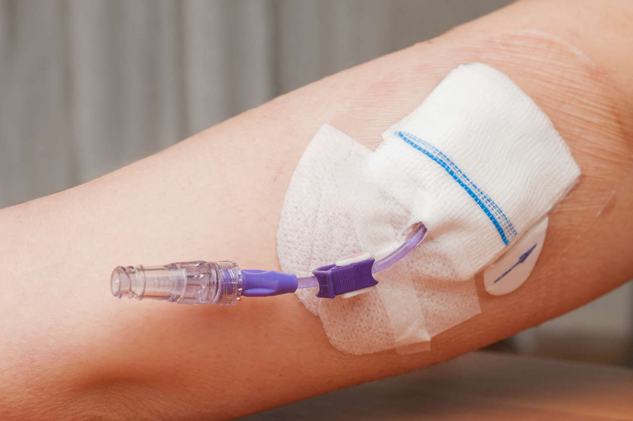
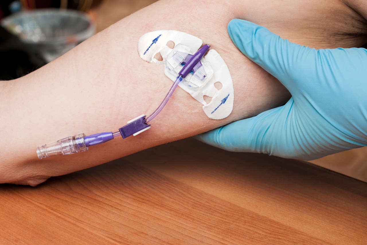
Tooth extraction is an invasive process. Depending on the disease severity and the number of teeth to be extracted, you may need replacement clotting factors before the procedure. 

After your tooth has been extracted, you should avoid rinsing, smoking, or strenuous activities for 24 hours. Also, you shouldn’t take pain medications that increase bleeding risk, such as aspirin. 

Fillings, Crowns, and Bridges

These restorative treatments are considered low-risk. Thus, they may be carried out in general dental practice. 

Root Canal Treatment

Root canal treatment doesn’t appear to cause additional bleeding problems in people with hemophilia. 

Get Financial Assistance

Schedule a Consultation


Hemophilia and Dental Treatment: Frequently Asked Questions

How do you stop gum bleeding from hemophilia?

Tranexamic mouthwash and tablets are effective in stopping gum bleeding in hemophilia. 

What happens if I need a tooth extraction? 

Tooth extraction is an invasive process. You may need replacement clotting factors or other medications before the procedure. You will also need to follow your dentist’s instructions after the procedure. 

What should I do if brushing makes my gums bleed?

First, talk to your dentist and follow their instructions, as it could be a sign of gum disease. Continue to brush your teeth (for two minutes per session) at least twice daily with fluoride-containing toothpaste. Use a brush with medium-texture bristles.

What To Do When IVIG Doesn’t Work: IVIG Alternative Treatments

Worried about what to do when intravenous immune globulin (IVIG) doesn’t work or fails to work as expected? Take a breath. You have several treatment options with comparable or even better outcomes. 

Highlights

  • When IVIG doesn’t work, your provider may recommend alternative treatments, such as plasmapheresis, corticosteroids, and immunosuppressive agents. 
  • The choice of an alternative treatment is highly individualized.
  • Alternative treatments depend on co-existing medical conditions, age, potential risks, and availability/cost.

IVIG is an effective treatment for numerous autoimmune, inflammatory, and infectious conditions. But, like all medications, not everyone responds to IVIG equally, and some may not respond at all. 

Several factors may determine the success or failure of IVIG therapy, such as:

  • Individual patient factors
  • Age 
  • Adherence to treatment plan 

Likewise, studies show that the percentage of responders can vary depending on the condition being treated. In this article, we discuss the alternatives to IVIG specific for various medical conditions. 

Also Read: How To Tell if IVIG Is Working

IVIG Alternatives in Chronic Inflammatory Demyelinating Polyneuropathy (CIDP)

Though IVIG is a first-line treatment for CIDP, about 1 in 4 people with CIDP don’t respond to it. When IVIG doesn’t work, your provider may prescribe immunosuppressive agents or monoclonal antibodies (mAbs) [1]. Additionally, some experts recommend switching from IVIG to SCIG (subcutaneous immunoglobulin).  

Immunosuppressive agents work by preventing the immune system from attacking the protective covering around nerve cells. Examples of immunosuppressives include azathioprine, mycophenolate mofetil, methotrexate, cyclosporine, and cyclophosphamide

Rituximab, a monoclonal antibody, improves CIDP symptoms in treatment-resistant cases according to a 2022 review [2].

IVIG Alternatives in Guillain Barre Syndrome (GBS)

IVIG is the primary treatment for GBS. Yet, about 25% of people who don’t respond to it need mechanical ventilation, and 20% of people cannot walk without support after 6 months [3].

When IVIG doesn’t work, your provider may switch you to plasmapheresis (plasma exchange) [4]. Plasmapheresis helps by removing the antibodies that attack your nerves. 

IVIG Alternatives in Kawasaki Disease (KD)

Though universal treatment guidelines are not available for KD, alternative treatments for IVIG-resistant KD include [4]:

  • A corticosteroid called methylprednisolone
  • A monoclonal antibody called infliximab (often considered the drug of choice)

IVIG Alternatives in Bleeding Disorders

If you have a bleeding disorder called immune thrombocytopenia (ITP), you are at risk of blood loss, especially during surgery. IVIG is commonly used before surgery to reduce the risk. 

A 2020 trial shows that an oral drug—eltrombopag—can be as effective as IVIG in people with ITP [5].

Consult an IVIG Specialist | The Best IVIG Home Infusion

(877) 778-0318


IVIG Alternatives in Immune Deficiency

Emerging evidence suggests that SCIG (subcutaneous immunoglobulin) may be a better option for people with immune deficiency. 
Unlike IVIG, SCIG results in the slow release of antibodies from the subcutaneous tissues into the blood. This allows antibody levels in the blood to remain steady. However, with IVIG, the antibody levels in the blood are more erratic.
Moreover, SCIG is more convenient because you can inject your dose at home or anywhere else at any time of day after some training. Additionally, side effects are less common with SCIG compared to IVIG. 
 

What Can Be Used Instead of IVIG?

In general, SCIG causes fewer side effects than IVIG. You may choose SCIG over IVIG if you:

  • Cannot tolerate IVIG side effects
  • Have poor venous access
  • Find SCIG more compatible with your lifestyle

Further Reading: SCIG vs. IVIG: Which Treatment Is Best for You?

What Is Advate (Antihemophilic Factor [Recombinant])?

Advate is an FDA-approved medicine used to treat children and adults with hemophilia A. In people with hemophilia A, the blood does not clot as it should. 


Speak to a Specialist About Copay Assistance

(877) 778-0318

What Is Advate?

Advate is a brand-name prescription medication. The active medication in this product is recombinant clotting factor VIII. You may also call it a recombinant antihemophilic factor. It belongs to a class of medicines called antihemophilic factors. The term “recombinant” means Advate is made with recombinant DNA technology. 
Recombinant clotting factors are not derived from blood. Therefore, Advate is not a blood product and does not carry the risk of spreading blood-borne viruses. 
People with hemophilia A have little or no clotting factor VIII in their blood. Clotting factor VIII helps your body form blood clots. Factor VIII deficiency can lead to spontaneous, uncontrolled internal bleeding, which can be fatal. 
The FDA initially approved Advate to control and prevent bleeding episodes in hemophilia A in 2003. 

How Does Advate Work?

This medicine works by replacing factor VIII in people with hemophilia A.   

How Is Advate Supplied and Used?

This medicine comes as a white to off-white powder for intravenous infusion (IV) in single-use vials. The following strengths are available in the United States:

  • 250 IU
  • 500 IU
  • 1,000 IU
  • 1,500 IU
  • 2,000 IU 
  • 3,000 IU 
  • 4,000 IU

An IU (International Unit) is the amount of a substance that has a specific biological effect (in this case, to help your blood clot). 
Before administering the drug, your provider will determine the dose. The dose and duration of treatment depend on:

  • Severity of factor VIII deficiency
  • Location and severity of bleeding
  • Your body weight and overall health
  • Desired factor VIII level
  • Condition being treated

Your provider will calculate the dose using specific formulas. Then, they will prepare the liquid for injection following instructions on the package. 
Your provider will administer the liquid over a period of 5 or fewer minutes. Before and during administration, you will have your pulse rate measured. If your pulse rate becomes high, they will likely reduce the rate of administration or temporarily stop the injection. Infusion should begin within 3 hours after dilution. 

What Is Advate Used to Treat?

Advate is used in children and adults with hemophilia A to:

  • Control and prevent bleeding episodes
  • Prevent bleeding during surgery
  • Reduce the frequency of bleeding episodes

This product is not used to treat a form of bleeding disorder called von Willebrand disease. 

What Are the Side Effects of Advate?

Woman suffering side effects from taking AdvateSide effects can be mild or severe. The most common side effects are:

  • Fever
  • Headache
  • Cough
  • Common cold
  • Joint pain
  • Vomiting
  • Upper airway infection
  • Stuffy nose
  • Diarrhea
  • Sore throat
  • Itching
  • Unusual taste
  • Swelling of legs
  • Chills 
  • Sweating
  • Rash
  • Fatigue

Talk to your provider if these side effects persist or worsen.  

Get Financial Assistance

Schedule a Consultation

 
Advate can cause severe side effects in some users. Anaphylaxis is a life-threatening allergic reaction that causes breathing problems and low blood pressure. Talk to your provider immediately if you develop the following symptoms after receiving Advate:

  • Rash and itching
  • Hoarseness
  • Breathing difficulty
  • Trouble swallowing
  • Swelling of your hands, face, or mouth

Some users may form molecules called inhibitors that may prevent Advate from working properly. If that happens, you will have problems stopping a bleeding episode. 
Those who develop inhibitors need special treatment until the body no longer makes inhibitors. Talk to your provider to learn more about inhibitors and blood tests to detect inhibitors. 
 

Who Shouldn’t Receive Advate?

People allergic to mice, hamsters, or any product component should not receive Advate. Inform your provider if you have any known allergies. 
 

Use in Pregnancy and Lactation

No human or animal data with use during pregnancy are available. Likewise, it is unknown if this medicine is present in breast milk or if it harms the breastfed infant. Tell your provider if you are pregnant or nursing a child.
 

Can You Self-Administer Advate?

If your provider has instructed you to self-administer this medicine, remember the following:

  • Use Advate exactly as directed. 
  • Follow all directions on the prescription label. 
  • Never use this medication in larger or smaller quantities or for longer than recommended. 
  • Be sure to check the strength of the medicine on the label. 
  • Do not self-administer if you are not sure how to use the injection.
  • Always use sterile water and a reconstitution device that comes with the product to prepare the solution for injection. 
  • Do not refrigerate the liquid after reconstitution. 
  • Use the liquid within 3 hours after reconstitution.
  • Before using your dose, visually check the solution. It should be clear and colorless. 
  • Discard liquid that is not clear or contains particles. 
  • A single-use vial, as the name suggests, is for one use only. 
  • Put the needle, syringe, and Advate in a puncture-proof sharps container before disposing. Do not dispose of any of them in ordinary household trash.
  • Keep the container away from children and pets.
  • You may need frequent blood tests to check if the treatment is working or if you need to change your doses. 


What Happens If I Miss a Dose?

Because this medicine is administered when needed, a dosing schedule is unlikely. However, if you are on a schedule, take the missed dose as soon as you remember. Skip the missed dose if it is time for the next scheduled dose. Never use extra medicine to compensate for the missed dose.
 

What Happens If I Overdose?

Seek emergency medical attention or call the poison help line at 1-800-222-1222.
 

What Is the Difference Between Advate and Adynovate?

Like Advate, Adynovate is used to help treat and control bleeding in people with hemophilia A. Both products contain the same active substance, recombinant clotting factor VIII. However, Adynovate stays longer in your system, allowing less frequent dosing. 
 

How Much Does Advate Cost?

The amount you pay for Advate treatment can vary, depending on your insurance plan, geographical location, and pharmacy. Contact your insurance provider to find out if your plan covers this medicine or if you need prior authorization.
According to Takeda Pharmaceuticals U.S.A., which makes Advate, over 90% of commercial and Medicaid health plans cover this medicine. 
If you are new to Advate treatment, you may also be eligible to get 6 free trial doses with Takeda’s FREEDOM OF CHOICE program. 
Contact us below to learn more about financial assistance for Advate. 
 

Kawasaki Disease: 10 Frequently Asked Questions, Answered

This article answers the 10 most common questions about Kawasaki disease. Empower yourself with evidence-based health information to make better choices. 

Highlights

  • Kawasaki disease (KD) is a rare condition that usually affects children younger than 5. Older children and adults rarely get KD. 
  • KD causes the blood vessels to become inflamed. Typical symptoms include fever, rash, and a red strawberry tongue. 
  • What causes Kawasaki disease is unknown. Researchers are studying the possible role of genetics and environmental factors, such as infections. 
  • Diagnostic criteria involve the presence of symptoms such as fever, rash, strawberry tongue, and others. 
  • Prompt treatment, with high-dose IVIG and aspirin, results in complete recovery in most children. 

What Is Kawasaki Disease?

Kawasaki Disease is also called Kawasaki syndrome or mucocutaneous lymph node syndrome.

Kawasaki disease is a form of vasculitis, which is inflammation of the blood vessels. Inflamed blood vessels cannot supply enough blood to tissues and organs. Moreover, they become weak and prone to tearing. 

KD causes swelling of the blood vessels throughout the body. But heart complications are most likely when blood vessels that supply the heart (coronary arteries) get affected. KD is one of the leading causes of heart disease in children in the US. 

KD isn’t contagious, though there have been reports of occasional community-wide outbreaks. 

How Common Is Kawasaki Disease?

According to the Centers for Disease Control and Prevention (CDC), KD affects about 9 to 20 per 100,000 children younger than 5 in the US. In contrast, 50 to 250 per 100,000 children in Japan, Taiwan, or Korea develop KD [1,2].

In 2016, out of 5,440 KD-related hospitalizations in children under 18, 3,935 involved children younger than 5.

What Causes Kawasaki Disease?

Experts are yet to identify what exactly causes Kawasaki disease. However, initial research suggests it might be linked to genes, viruses, bacteria, and environmental triggers like irritants. 

Recent studies show that carrying specific genetic changes may make your child more likely to get KD. Having a sibling with KD may increase the risk severalfold. In addition, over 4 in 10 children diagnosed with KD have a viral respiratory infection [3].

Who Gets Kawasaki Disease?

Kawasaki disease occurs most often in children between 6 months and 5 years of age. Older children, children younger than 4 months, and adults rarely get KD. 

In a 2015 case report, Japanese researchers reported a very rare case of KD in a 37-year-old Japanese man [4]. Talk to your doctor if you have a fever with no known cause and rash, especially if you have a current or recent history of a viral infection. 

Other factors that may increase the risk of KD include:

  • Sex: Boys are more likely than girls to develop KD and its complications, including death. 
  • Ethnicity: Children of Asian descent, particularly Japanese, are at the highest risk. 
  • Seasonality: KD is more likely to occur in the winter and spring months. 

Kawasaki Disease and COVID-19: Is There a Link?

Available studies haven’t found a link between KD and COVID-19. 

In some children, COVID-19 may cause a condition with symptoms that resemble KD. Doctors call this condition multisystem inflammatory syndrome in children (MIS-C). 

MIS-C may also cause additional symptoms, such as:

  • Abdominal pain
  • Nausea
  • Vomiting
  • Diarrhea
  • Headaches
  • Low blood pressure

While Kawasaki disease typically occurs in children, MIS-C can affect both children and adolescents.

What Are the Symptoms of Kawasaki Disease?

Boy with fever from Kawasaki disease

Kawasaki disease occurs suddenly, and symptoms appear in phases. 

Phase 1 Symptoms

  • High fever (over 101°F) that lasts more than 5 days and doesn’t improve with medications
  • Rash on the trunk and in the genital area
  • Bloodshot eyes with no thick discharge
  • Strawberry tongue
  • Red, swollen skin on the palms and soles
  • Swollen lymph nodes in the neck

Phase 2 Symptoms

Phase 2 symptoms may include:

  • Skin peeling on the tips of the fingers and toes
  • Joint pain
  • Diarrhea
  • Vomiting
  • Abdominal pain

Phase 3 Symptoms

During the third phase, the signs and symptoms will go away slowly. It might last up to 8 weeks. Your child may show symptoms of tiredness, irritability, and low energy. 

How Is KD Diagnosed?

No specific diagnostic test for KD exists. Your child’s doctor will make a diagnosis based on the symptoms and physical exam. 

The diagnostic criteria for KD is a fever lasting over 5 days and having at least 4 of the following 5 features [5]:

  1. Strawberry tongue
  2. Rash
  3. Bloodshot eyes with no thick discharge
  4. Swollen, red skin on the palms of the hands and the soles of the feet
  5. Swollen lymph nodes in the neck

Incomplete or atypical KD is when children don’t fulfill the usual criteria for KD diagnosis but still have the disease. 

How Is Kawasaki Disease Treated?

Prompt treatment is crucial to reduce the risk of complications. Children who stay in the hospital for treatment usually receive:

Aspirin

Your child will receive moderate to high dose aspirin until fever subsides. Then, a low dose is given for up to 8 weeks. 

IVIG

Timely treatment with IVIG can greatly lower the risk of heart complications. IVIG contains proteins called antibodies. These proteins can stop your immune system from attacking the blood vessels. 

Corticosteroids

Corticosteroids may be an option for children who don’t respond to IVIG. 

What Are the Complications of Kawasaki Disease?

Potential complications include:

  • Aneurysms (bulging in the wall of a blood vessel)
  • Abnormal heart rhythm
  • Blood clots (may lead to heart attacks)
  • Swelling of the coronary arteries
  • Heart valve problems

What Is the Life Expectancy for Someone with KD?

If blood vessels in the heart aren’t affected, full recovery is possible. Heart attack due to aneurysms is the main cause of death.

Lifelong monitoring is necessary in children with aneurysms or other heart complications. 

According to a 2022 review, survival ranges from 92% to 99% at 10 years and 85% to 99% at 20 years [6].

Get Financial Assistance

(877) 778-0318

Myasthenia Gravis Causes: Factors That Contribute to This Condition

According to the Myasthenia Gravis Foundation of America, around 36,000 to 70,000 people have been diagnosed with myasthenia gravis (MG) in the United States alone. 

Speak to a Specialist About Copay Assistance

Schedule a Consultation

 
Myasthenia gravis is an autoimmune neurological disorder that causes severe muscle weakness, which can later lead to respiratory failure as the muscles that function to keep the airway open are compromised. 

Though the exact cause that triggers this neurological condition remains unclear, it is believed that a combination of genetic, immunological, environmental, and physiological factors is involved in the onset of myasthenia gravis. 

Below, we have listed some myasthenia gravis causes that are believed to contribute to the onset of this condition.

Myasthenia Gravis Causes

Several common factors, like age, thymus gland irregularities, immune attack, infection (viral or bacterial), immunization, or rarely surgery, can trigger the onset of myasthenia gravis. However, among all factors, immune dysfunction plays a significant role in the onset and progression of myasthenia gravis. 

Let’s discuss the mechanism through which the immune system and other factors trigger this neurological condition. 

Immune System Dysfunction

Under normal conditions, the immune system works to protect your body from harmful pathogens by producing antibodies that detect and kill them. However, when the immune system becomes dysfunctional, it starts attacking your healthy cells, mistaking them for these foreign pathogens. 

In myasthenia gravis, the neuromuscular junction (an area where the nerve and muscle connect and communicate) is attacked by the immune system. In a healthy person, acetylcholine binds to the acetylcholine receptor (AChR) at this junction of muscle cells which subsequently causes muscle contraction. However, in MG, the immune system produces autoantibodies that block the acetylcholine receptor protein. 

When the autoantibodies bind to the acetylcholine receptors (AChR), the ability of the receptor to receive the chemical signal, acetylcholine, is reduced. As a result, due to insufficient or low chemical signals, muscles do not contract. Hence, people with MG experience extreme fatigue and profound muscle weakness.

Thymus Gland Abnormalities

Male thymus gland

Abnormalities in the thymus gland have also been associated with MG. Normally, the thymus gland regulates the development of T-cells (a type of immune cell) and trains them to distinguish the healthy body’s cells from harmful pathogens. This gland grows gradually until puberty, then shrinks and is replaced by fats. 

Sometimes, thymus glands remain large even after puberty and transform into cancerous cells. Researchers believe that it is possible that when the thymus gland does not work properly, the developing T-cells lose their ability to distinguish between healthy and harmful cells. As a result, they attack the body’s healthy cells and tissue. 

For example, in 15% of cases, people with MG have a thymic tumor (or thymoma), and in 75% of cases, MG people have thymic abnormalities (thymic hyperplasia).

Age

Age is a risk factor for MG. Young women in their 30s and older men in their 60s and 70s are most likely to develop MG. It is more common in adults; however, MG can be diagnosed in children.

Viral or Bacterial Infection

Viral or bacterial infection may also trigger or contribute to the onset of myasthenia gravis symptoms. Sometimes, bacterial or viral proteins mimic the structure of acetylcholine receptor protein (AChR), which stimulates the immune system to attack the healthy AChR protein. 

For instance, in a case report, three patients with no previous record of neurological condition showed symptoms of myasthenia gravis within 5 – 7 days after the onset of COVID-19 infection. Researchers suspected that antibodies produced against COVID-19 proteins may cross-react with acetylcholine receptor subunits, which results in the onset of MG. 

Get Financial Assistance

(877) 778-0318

Genetics

Though MG is not a hereditary disease, it is possible that if an individual has any autoimmune conditions like rheumatoid arthritis or hyperthyroidism, they may also be susceptible to developing MG. However, the research on the association of autoimmune conditions with MG is still ongoing. 
 

Conclusion

Myasthenia gravis or MG is a complex neurological condition that can be caused by multiple factors, including immune system dysfunction, thymus gland abnormality, age, infection, or genetics. However, further research is needed to understand how each factor can cause myasthenia gravis in order to develop effective treatment strategies. 
 

TPN Through PICC Lines: What You Need to Know

If your TPN (total parenteral nutrition) therapy is expected to last up to several weeks, such as for 2 to 6 weeks, you will likely get a PICC line placed instead of a regular IV line. 

Your IV Fluids, Always Available

Full inventory, hassle-free access
(877) 778-0318

A PICC line, also known as a peripherally inserted central catheter, is a long, super thin, flexible tube (catheter) that is inserted into one of the peripheral veins (such as the basilic or cephalic vein) of your upper arm. The tube is then advanced through a larger blood vessel that leads to your heart, such as the subclavian vein or superior vena cava, typically present just above the right side of your heart. 

The PICC line placement procedure is mainly done by a registered nurse or physician assistant using a sterile technique.

Before undergoing TPN treatment through PICC lines, it is important to know why it’s considered a preferred choice for TPN infusion, how it is placed, and what risks or complications you can expect during TPN or after TPN infusion 

Why Is the PICC Line Used for TPN Therapy Instead of Regular IVs or Other Central Lines?

There are a number of reasons that healthcare providers consider providing TPN infusion through a PICC line instead of regular intravenous lines or central lines:

It Provides Long-Term Access

The PICC line is designed in such a way that it can stay in place for several weeks or even months. This long-term access makes it easier for patients to receive TPN therapy for up to a few months without frequent reinsertion. 

Furthermore, unlike a regular intravenous (IV) line, which is shorter, the PICC line is a super thin, long tube that provides easy access to the larger vein near the heart. 

It Reduces Discomfort

The PICC line reduces the discomfort caused by repeated needle sticks with general IV lines and can deliver large amounts of IV fluids, which might not be possible through regular IV lines. 

It Minimizes the Risk of Infections

PICC lines are considered safe and cost-effective because they have a lower infection risk than other central lines and regular IV lines.  

How Is TPN Given Through the PICC Line?

To provide TPN infusion through a PICC line, the healthcare provider will first place a PICC line. 

PICC Line Placement Procedure

TPN solution hanging from stand

The PICC line insertion procedure takes about an hour and is typically done in a general room equipped with imaging technology, such as X-ray machines. To insert a PICC line, a healthcare provider will do the following steps:

  • Clean the insertion site and inject the numbing agent, such as 1% lidocaine, at the insertion site in your upper arm. The numbing agent reduces the pain and discomfort during incision. 
  • Once the numbing agent is injected, the doctor will carefully make a small incision to access the vein and put the PICC line into the vein.
  • The PICC line is gently threaded into the vessels and advanced toward the heart using an ultrasound.
  • A chest X-ray will be performed to ensure the PICC line is correctly positioned. 
  • Once the line is confirmed with an X-ray, your doctor will then secure the line with a bandage or stitches. 
  • The two or three tubes sticking out of your skin, called lumens, will be used to deliver TPN solution to your body. 

Infusion of TPN Through PICC Line

Once the insertion is successfully completed, your doctor will start the TPN infusion procedure. To learn how to start the TPN infusion process, read our article: A Complete Step-by-Step Guide To Administering TPN. 

Ask About TPN Home Infusion

Schedule a Consultation

How Do You Care for and Maintain Your PICC Line After the TPN Infusion?

Once your TPN infusion process is complete, you need to secure and maintain the line carefully. 

  • Always flush and lock the PICC line with saline and heparin solution after TPN infusion. Your healthcare provider or nurse will give you directions about cleaning and flushing the line weekly to prevent blockage. 
  • Check your insertion site daily for signs of infection, swelling, or redness. If you see redness or feel pain around the insertion site, consult your healthcare provider immediately. 
  • Avoid lifting heavy weights, as doing so can affect your line.
  • Do not use the affected arm to check blood pressure.
  • Cover your PICC line with plastic wrap or waterproof bandage when taking a shower.
  • Do not change the dressing of your PICC line by yourself; instead, ask your nurse or trained caregiver to put the dressing over it.

Are There Any Risks Associated With TPN Given Through a PICC Line?

No particular risks are associated with TPN given through a PICC line compared to other modes of TPN delivery, as long as the line is secured and maintained properly. However, the PICC line can sometimes become clogged with medication or blood clots if not flushed properly with saline and heparin solution after the TPN infusion. 

If the line gets clogged, the TPN fluid may not flow smoothly. This requires the removal and replacement of the line with a new PICC line. 

When To Consult Your Healthcare Provider

Consult your healthcare provider immediately if:

  • You have a high fever or experience shortness of breath, itching, or dizziness.
  • Your line won’t flush or is blocked.
  • Your PICC line is leaking.
  • You have bleeding from the PICC line site.
  • Your PICC line accidentally comes out.
  • Your PICC line dressing becomes loose or dirty.

How Long Does IVIG Take To Work? Timeline & What To Expect

You will most likely experience improvements in your symptoms within several days to weeks after receiving IVIG therapy


Treatment Info | Get IVIG Prior Authorization

(877) 778-0318
 

What Is IVIG?

IVIG, or intravenous immunoglobulin, is a medication containing antibodies. Antibodies are proteins that help you fight against infections. IVIG is typically given as an infusion through your vein (intravenously), but some brands can also be given as an injection under the skin (subcutaneously).  IVIG is commonly used to treat certain diseases of the nerves and muscles, including:  

How Long Does IVIG Take To Work?

It is difficult to know for sure. However, experience and research show that most people can start to feel better within a few days to weeks after the first treatment. However, not everyone responds to IVIG treatment. People are categorized into two types: Responders and nonresponders.  One study shows that 6 in 10 people with chronic inflammatory demyelinating polyneuropathy respond to IVIG treatment [1]. On the other hand, 8 in 10 people with immune thrombocytopenia (ITP) respond to IVIG [2].  In those who respond, the time it takes for IVIG to work depends on several factors, such as:

Specific IVIG Brand

IVIG products are unique and not interchangeable. Each product has its own unique properties, which affect how it is absorbed, processed, and removed from the body. Consequently, these factors determine how long IVIG takes to work. 

Condition Being Treated

The condition you are taking IVIG for also determines how long IVIG takes to work. For example, IVIG raises the platelet count in immune thrombocytopenia within 4 days. But some people may respond within 24 hours [2]. On the other hand, for myasthenia gravis, IVIG can take up to 4 weeks to show its effects [3].

Patient Factors

Because each person is different, their response to IVIG or any other treatment varies greatly.  Their general well-being and the presence of certain diseases, particularly liver and kidney disorders, determine how soon their symptoms improve. 

Concomitant Use of Other Medications

How soon you can expect to feel better may depend on whether you are taking IVIG alone or with other medications. One medication can affect how your body processes the other, which can affect how quickly or how well IVIG works.   

Speak to a Specialist About Copay Assistance

Schedule a Consultation
 

How Can You Tell if IVIG Is Working?

3D illustration of human antibodies While subjective experience of well-being is a strong indicator to tell if IVIG is working, several other objective indicators do the same, perhaps in a more reliable way. These include:
  • Disease symptom score
  • Number of relapses in a particular period 
  • Laboratory findings
  • Frequency of flare-ups
 

What Is the Response Rate of IVIG?

The response rate of IVIG can vary depending on a specific disease or dose. For example, the response rate is 60% in chronic inflammatory demyelinating polyneuropathy (CIDP) and 80% in people with immune thrombocytopenia (ITP).   

How Often Should IVIG Be Given?

IVIG doses are typically given every 21 to 28 days (3 to 4 weeks). Sometimes, a single dose may suffice. However, your healthcare provider will ultimately decide how often you should receive IVIG based on your condition and symptoms.  

Why Is Hemophilia More Common in Males?

Hemophilia is more common in males due to the fact that they have only one copy of the gene on the X chromosome. If any changes occur in that gene, females can compensate for the defect with an additional copy of the gene, but males cannot. 

Speak to a Specialist About Copay Assistance

Schedule a Consultation

It is also the reason why more males get other X-linked disorders like color blindness. X-linked disorders occur due to changes in the gene on the X chromosome.  

A Quick Overview of Hemophilia, Causes, Types, and Occurrence

Hemophilia is a genetic disorder in which the blood does not clot as it should. As a result, it can lead to excessive bruising and nosebleeds. Severe hemophilia can cause spontaneous bleeding (bleeding with no obvious cause). 
Hemophilia occurs due to changes in the genes that instruct the body to make blood-clotting proteins called clotting factors. The common types are:

  • Hemophilia A: Also known as classic hemophilia, it occurs due to having little or no clotting factor VIII. 
  • Hemophilia B: Also known as Christmas disease, it occurs due to having little or no clotting factor IX. 

The more common type—Hemophilia A—affects 1 in 5,000 male live births. According to reports from the Centers for Disease Control and Prevention (CDC), nearly 400 babies are born with this disorder each year in the U.S. Though the exact numbers remain unknown, recent data suggest about 33,000 American males have this condition [1].
 

Why Hemophilia Is More Common in Males: Understanding the Genetics and Inheritance

The fundamental reason why hemophilia is more common in males is that they have only one X chromosome. 

The X chromosome contains the genes for clotting factors VIII and IX. Changes in these genes lead to little or no clotting factor production. Consequently, the blood does not clot properly. 

The X chromosome contains the genes for clotting factors VIII and IX. Changes in these genes lead to little or no clotting factor production. Consequently, the blood does not clot properly. 

Because males have only one chromosome, one altered copy of the gene is enough to cause symptoms. On the other hand, females have two copies of these genes, and changes in one copy typically do not cause the condition. 

Moreover, males get an earlier diagnosis than females. Early diagnosis in males is a result of the myth that females cannot get hemophilia. Many females may even ignore the signs of mild hemophilia, such as heavy bleeding. 

According to a 2020 study, females with moderate symptoms receive a diagnosis about 6 months later than males. In severe cases, females face delays of approximately 40 months [2].

Hemophilia is more common in males born to a female carrying the affected gene and an unaffected male. In such cases, there is a 1 in 4 chance of having a baby boy with the condition. On the other hand, males born to a hemophilic father and unaffected mother are less likely to get the condition. 

The risk of hemophilia is the same for males and females if both parents have the faulty gene. In such cases, there is a 1 in 4 chance that a baby girl will be a carrier. 

Get Financial Assistance

(877) 778-0318

Why Is Hemophilia Rare in Females?

For females to get hemophilia, both X chromosomes have to be altered. Sometimes, hemophilia may result from having one affected X chromosome and another non-functioning chromosome.  

A female with one altered gene is a “carrier.” She can pass the affected gene on to her children. Female carriers do not usually experience symptoms of hemophilia, but some do. 

The prevailing misbelief that hemophilia is a “male-only” disease could be one of the reasons for fewer diagnoses in females.

Key Takeaways

  • Hemophilia is more common in males because they have only one X chromosome. 
  • Though some providers consider it a “male-only” disease, hemophilia can affect females as well.  
  • Many health experts think underrecognition and underdiagnosis in females could obscure the real-world scenario. Females appear to get delayed diagnoses. 

Central Line TPN Guidelines for Healthcare Professionals and Patients

If you or your loved one requires total parenteral nutrition or TPN for a long period of time, your healthcare provider will order TPN nutrients to be administered through a central line, also known as the central venous catheter (CVC), into your body. 

IV Fluids Fully in Stock

Reliable supply for uninterrupted care
(877) 778-0318

A central line or central venous catheter is a soft, thin, and flexible hollow tube that is inserted in a large vein, usually the veins located near the neck or chest. Unlike a regular IV tube, a central line tube is much longer and can stay in place for a month to several years. Furthermore, large amounts of fluids can be delivered easily through a central line, which would not be possible through peripheral catheters. 

In this article, we will discuss the main types of central lines as well as some guidelines that you can follow prior to or while receiving TPN nutrition through a central line. 

Types of TPN Central Lines

Central lines or central venous catheters are categorized into external central lines and internal central lines. External central lines include peripherally inserted central catheter (PICC) and tunneled CVC, while internal central lines include implanted ports. Each type is unique and has different insertion points. 

External Central Line

In a peripherally inserted central catheter (PICC) or tunneled CVC, a catheter is inserted in one of the veins of the upper arm (such as a basilic, brachial, cephalic, or medial cubital vein) or chest. This line is further advanced into a large vein above the right side of the heart (superior vena cava). The procedure of inserting a catheter is done by a registered nurse, doctor, or trained healthcare professional.

The other end of the line branches out to one or two smaller tubes called lumens, which, when connected to an infusion tubing, are used to deliver essential nutrients from the catheter directly into the vein. 

Internal Central Line

In an implanted port, a catheter is surgically inserted completely under the skin by a trained physician. This central line is usually placed in the chest and may sometimes be placed in the upper arm. 

The one end of the implanted port’s tube goes into the large vein, while the other end leads to a small rubber disc under the skin. Nutrients are administered with the help of a special type of needle called a Huber needle, which is inserted through the skin into the port reservoir. 

Unlike the external central line, where the catheter tube sticks out the skin, in the internal central line, the catheter does not stick out of the skin except for a small bump you’ll feel under the skin.  

Central Line TPN Guidelines

Prior to TPN therapy, your healthcare provider will educate you about central lines, the type of central line you have, and how you can inject nutrients as well as maintain infusions at home. 

TPN Central Line Care and Maintenance

Before and after TPN infusion, it is important to protect your central line catheter from infection when not in use. In most cases, the central line lumen is flushed (clean) with a saline solution. This helps to remove medication residues that may have been left during the last infusion and to prevent the risk of blockage during your TPN infusions. 

Flushing the Central Line Lumens

Healthcare worker helping patient set up TPN
  1. Gather your supplies, including gloves, alcohol pads, aseptic caps, saline and heparin pre-filled syringes. 
  2. Wash your hands and put on gloves.
  3. Remove the end cap and clean the outside of the lumen with an alcohol pad for 15 seconds in a twisting motion. Let it dry for 5 seconds. 
  4. Attach the pre-filled saline syringe to the lumen by pushing it onto the lumen and turning it clockwise to lock it in place. 
  5. Release the clamp on the lumen of your CVC and slowly start to flush the lumen by pushing down the plunger slowly. 
  6. Push about 1 – 2 ml of saline at a time using the push and pause method until all the saline is used from the syringe.
  7. Clamp the lumen back and unscrew the saline syringe in a counter-clockwise motion.
  8. After flushing the central line, carefully attach the IV tubing to your catheter by using a push and twisting motion to secure the connection. To learn how to start the TPN infusion process, read our following article: A Complete Step-by-Step Guide for Administering TPN.
  9. Once the TPN infusion process is complete, clamp your lumen back and carefully disconnect the IV tubing from the catheter.
  10. Perform the post-flushing of the lumen by following steps 3 – 7 above and cover the lumen ends with aseptic caps. 

Speak to a Specialist About Copay Assistance

Schedule a Consultation

Locking the Central Line Lumens

The method of locking the central line is similar to flushing the lumen; you will only use heparin instead of saline. The purpose of locking the central line is to prevent the formation of blood clots in the catheter and at the catheter tip. It also helps to prevent microorganism adhesion and biofilm formation.

  1. Attach the pre-filled heparin syringe with the lumen by pushing it onto the lumen and turning it clockwise to lock it onto the lumen. 
  2. Now, release the clamp on the lumen of your CVC and slowly start to push the plunger. 
  3. Push 0.5 ml of heparin at a time using the push and pause method until all 3 ml of heparin is used.
  4. Clamp the lumen while keeping your thumb on the plunger, unscrew it, and remove the heparin syringe carefully.
  5. Cover the lumen end with the aseptic cap.

Repeat the flushing and locking steps with the saline and heparin solution on the other lumens if you have more than one. 

Flushing and Locking of Implanted Port Central Line

You can apply an anesthetic cream 1 to 4 hours before infusion to reduce the discomfort of needle insertion. Over time, the skin over the port thickens, making it less sensitive to needle sticks.

Cleaning the Port Site

  1. Sterilize the skin (catheter site) with an aseptic solution for 30 seconds back and forth, up and down, sideways, and in a diagonal pattern.
  2. Allow the port site to dry for 30 seconds. 

Access the Implanted Venous Device

  1. Use one hand to stabilize the border of a venous access device and use the other hand to pick up the Huber needle with a syringe attached.
  2. Grasp the wings of the safety needle with your thumb and middle finger. 
  3. Insert the needle carefully into the septum (silicone top) at a 90-degree angle perpendicular to the skin surface and advance the needle through the skin and septum until it makes contact with the bottom of the port reservoir. 
  4. Once you’ve successfully placed the needle, remove the protective cap from the needle wings, stabilize the needle wings, and apply an antimicrobial patch around the needle.
  5. Now, place a sterile dressing (bandage) on the skin covering the port to lower infection risk.
  6. Administer a 10 ml saline solution by slowly pushing down the plunger. 
  7. Push about 1 – 2 ml of saline at a time using the push and pause method until all the saline is used from the syringe.
  8. While injecting, observe skin around the Huber needle site for leakage of fluid or infiltration to access the device. 
  9. Once you’ve injected the saline solution, gently remove the syringe from the end cap and clamp the tubing. 
  10. If you’re giving a TPN infusion, leave the needle in place for the infusion. To learn how to start the TPN infusion process, read the following article: A Complete Step-by-Step Guide for Administering TPN.
  11. After infusion, perform post-flushing with normal saline (repeat from steps 1 – 9).

Locking Implanted Port Central Line Lumen

  1. Attach the pre-filled heparin syringe tip to the injection cap and flush using the push-and-pause method. When the syringe is empty, remove it from the end cap.
  2. Clamp the IV tubing.

De-accessing (Removing) Port Needle

  1. If you want to remove a needle from the port, hold the port steady with the fingers of one hand and gently raise the needle trap at a 90-degree angle with the other hand.
  2. Grasp the needle stick guard and hold it down firmly. 
  3. While holding the stick guard firmly in place, grasp the flexible wings and pull upward until the needle is completely encapsulated in the needle trap. 
  4. Cover the site with a bandage. 

Central Line TPN Guidelines: When to Consult a Doctor

Consult your healthcare provider immediately if you have any of the following issues:

  • Infection, swelling, or redness around the insertion site 
  • High fever
  • Problems during flushing of the lumens
  • Side effects like cardiac arrest, pneumothorax, or catheter-related bloodstream infections

How Is IVIG Made? The Intricate Steps of IVIG Manufacturing Revealed

Intravenous immunoglobulin (IVIG) is a therapeutic product prepared from the pooled plasma of thousands of donors. Human plasma contains several essential components, including immunoglobulins (Ig), also known as antibodies. Immunoglobulins are important components of the immune system and help the body fight infections. 

Ask About IVIG Home Infusion

(877) 778-0318

The process of purifying immunoglobulin from plasma to create patient therapies is complex and time-intensive. It takes approximately 9 months to produce a batch of IVIG products for therapeutic use.

The final IVIG product consists of a mixture of antibodies that are administered intravenously into the patient’s bloodstream. 

In this article, we will discuss the series of steps that every manufacturer follows to produce a final sterile solution of IVIG for therapeutic use. 

IVIG Production Process

The steps involved in the production of IVIG include plasma collection, fractionation, purification, formulation, testing, and packaging. 

Collection of Plasma

The manufacturing process of IVIG begins with the collection of plasma from thousands of healthy donors. Donors are selected through rigorous screening to ensure the highest quality and safety of plasma. 

Plasma used for IVIG production is obtained from two sources: About 20% comes from blood donors, while the remaining 80% comes from plasma donors.

Since immunoglobulins are a small part of plasma, many donations are required to produce enough IVIG to treat patients. For example, producing enough IG to treat one immune-deficient patient for a year takes about 130 donations from healthy individuals.

Plasma Fractionation

After blood plasma collection, a series of fractionation processes take place, during which plasma proteins such as immunoglobulins, clotting factors, and albumin are separated from other plasma components. 

Cohn’s fractionation technique, developed by Edwin J. Cohn in World War II, is typically followed, with some newer modifications, to extract a concentrated immunoglobulin solution from plasma. In this technique, small amounts of alcohol (ethanol) are used to reduce pH and ionic strength in order to separate albumin and other plasma proteins based on their solubility. 

During the process, the temperature is kept low to decrease the possibility of contamination and allow large-scale fractionation.

Purification of IVIG

IVIG fluid being administered in hospital

Once the immunoglobulins are separated via fractionation methods, rigorous purification ensues to produce a concentrated immunoglobulins solution. The purification process removes inactive proteins, impurities, and contaminants from the solution. For the purification of immunoglobulins, different chromatography methods, such as ion exchange chromatography using DEAE Sephadex®, hydrophobic charge induction chromatography, and size exclusion chromatography, are followed. 

Each chromatography technique is unique and yields 95% – 97% of highly purified IgG from small volumes of human plasma in a one-step purification process. For instance, DEAE Sephadex® column chromatography produces a highly purified 95% IgG solution. 

In contrast, size exclusion chromatography helps to separate the different forms of IgG immunoglobulin based on their respective size from the pooled plasma. 

Furthermore, chromatographic procedures also help to remove an IgA immunoglobulin, which can cause severe anaphylactic shock in patients with IgA deficiency and have anti-IgA antibodies in their blood.

Removal of Harmful Pathogens

Since IVIG is composed of plasma pools from thousands of donors, there is a chance that a virus such as HIV or Hepatitis C may be contained in a sample. Although much care is taken when choosing IVIG donors by completing donor screening and checking donated samples for viruses or bacteria, the use of a fractionation process is vital in obtaining a high level of purity in a sample.

Furthermore, several methods are designed to remove or inactivate the viral pathogens, as follows:

  • Solvent/detergent (S/D) treatment inactivates viruses by applying chemicals that destroy their outer coating (envelope).
  • Pasteurization is a process in which IVIG solution is heated to 60 ℃ for 10 hours.
  • UVC radiation and low-dose radiation methods are used to destroy the virus by attacking its genetic makeup. 
  • Low pH treatment disrupts the protective layer of the virus and changes protein conformation. 
  • Nanofiltration is a type of ultrafiltration method in which an IVIG solution passes through a membrane (filter) whose holes are too small for a virus to fit through. 

Can IVIG help? | Free IVIG Treatment Info

Schedule a Consultation

Formulation and Composition of IVIG

Once IVIG is purified, it undergoes a formulation and stabilization process to ensure its efficacy and stability. Manufacturers formulate and stabilize the IVIG solution by adjusting the pH and adding stabilizing agents such as sugar or amino acids. This step makes each IVIG product differ in terms of formulation and composition. 

Final Testing and Packaging of IVIG

IVIG products undergo stringent testing and quality control procedures throughout the production process. A variety of tests and assays are conducted to remove the pollutants, viral pathogens, or prions. 

Once the quality control tests are completed, the final product is transferred to vials under an aseptic environment and incubated for several weeks to ensure high quality and safety. 

After the final inspection, IVIG is labeled, packaged, and delivered to healthcare facilities for therapeutic use. 

Conclusion

IVIG is made from the pooled plasma of thousands of donors, which is then purified through rigorous purification and quality control methods. From the collection of plasma to the formulation of sterile IVIG solution, good collection practices are followed at every step to ensure the product’s safety, efficacy, and quality.