Tattoos are a form of body art that symbolizes personal stories, beliefs, important memories, emotions, or life experiences for the person wearing them. Nowadays, getting a tattoo in the U.S. is common and generally considered safe for people with normal clotting function.
However, for individuals with hemophilia, getting a tattoo may pose additional risks and challenges. Even though medical providers often discourage people with bleeding disorders from getting a tattoo or piercing, people may still decide to get them anyway.
According to HemAware (The Bleeding Disorders Magazine), people with hemophilia can safely get a tattoo by taking appropriate precautions. This article highlights the potential risks of tattoos for hemophilia patients and what precautions they can take to avoid complications.
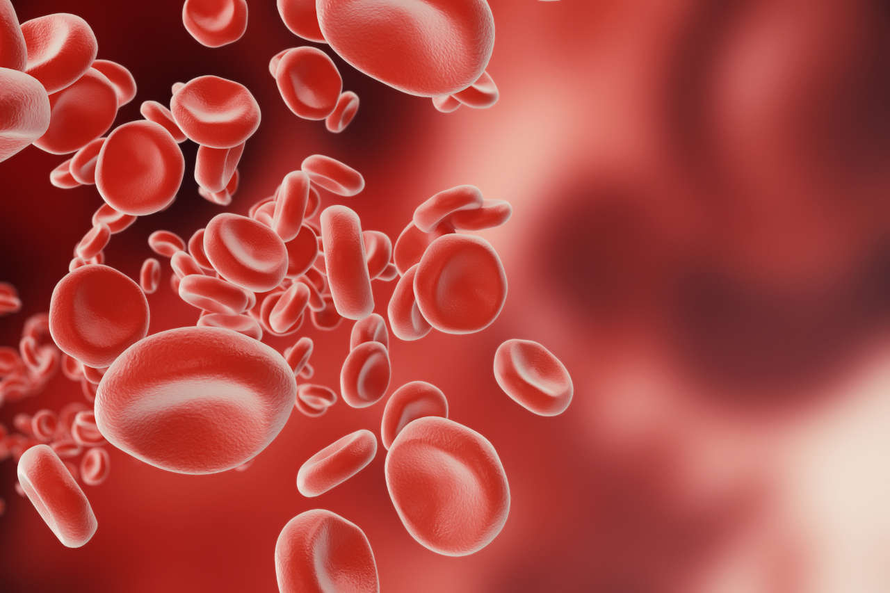
Hemophilia is a rare genetic bleeding disorder. In this disease, minor injuries can become potentially dangerous for a person with hemophilia due to a lack of clotting factors in their body.
Similarly, tattoos can also raise the chances of infection and significant bleeding in these individuals since the procedure used to make tattoos creates open wounds when the needle repeatedly punctures the skin to insert ink.
What Are the Possible Risks of Tattoos for People With Hemophilia?
If you have hemophilia, you should know the risks and concerns associated with tattooing. Some of them are as follows:
Excessive Bleeding
Anyone can experience bleeding while getting a tattoo. But, if you have hemophilia, you can bleed longer due to a lack of clotting factors. Excessive and prolonged bleeding is the biggest concern for hemophilia patients and, in severe cases, makes it difficult for the tattoo artist to continue and finish the process.
Bruising and Hematomas
Since it is difficult for hemophilia patients to form clots, bleeding under the skin can cause significant bruising and hematomas. This can slow down the healing process and affect the appearance of the tattoo.
Risk of Infection
The risk of infection also increases in hemophilia patients after getting a tattoo because the decreased ability to clot slows down the healing process. As a result, wounds created by tattoo needles take longer to heal.
Furthermore, the U.S. Food and Drug Administration (FDA) has reported cases of ink contaminated with bacteria or mold that can cause infections. If you get a tattoo in an unhygienic environment, your chance of getting an infection increases.
Allergic Reactions and Keloids
Some people with hemophilia may become sensitive to the pigments in the ink and experience allergic reactions during or after the tattoo procedure. If you have sensitive skin, you may also develop dermatitis, a form of skin irritation.
Similarly, some hemophilia patients may develop raised scar tissue near the tattoo, called keloids. Hence, it is recommended that you weigh the potential risks and benefits before getting a tattoo.
What Precautions Can You Take If You Have Hemophilia and Want a Tattoo?
Here are some precautionary steps that can help you get through the tattooing process as safely as possible:
1. Consult With a Medical Professional
If you have decided to get a tattoo, always consult with your medical professional, especially a hematologist. They will provide guidance about your current condition by assessing the severity of your hemophilia and advising you on whether getting a tattoo is safe or not.
2. Pre-Treatment With Clotting Factors
Always get pre-treatment with clotting factors before your tattoo appointment. This will help your blood clot normally and reduce the risks of excessive bleeding during and after the tattooing process.
It is recommended that an infusion be taken an hour before the tattoo procedure to maintain optimal factor levels.
3. Research and Choose an Experienced Tattoo Artist
Not all tattoo artists have experience in working with hemophilia clients and managing bleeding incidents during the tattooing process. Therefore, you should carefully look for an expert tattoo artist who uses sterile equipment and disposable needles and follows strict cleanliness procedures to minimize the risk of infection.
4. Choose Simple and Small Designs
If this is your first time getting a tattoo, consider choosing a simple and small design. This will limit the time you spend getting the tattoo and reduce the risk of bleeding. Additionally, smaller tattoos take less time to heal, and bleeding can be more easily managed.
The steps you take after getting your tattoo are just as important as the ones you take beforehand. Once you get a tattoo, you should follow the appropriate aftercare instructions to minimize the risk of infections or irritations. For example:
Use water or saline solution to clean the area carefully twice a day.
Avoid rubbing or scrubbing the tattoo area.
Avoid swimming and sun exposure until your tattoo has healed completely.
Moisturize the tattoo area several times a day.
Contact your doctor if you have an allergic reaction, swelling, or redness in the tattoo area.
Conclusion
Hemophilia patients can safely get tattoos with careful planning and consultation. However, the risks will vary depending on their disease severity.
Hemophilia is a rare genetic disorder that affects your body’s ability to form blood clots. As a result, you experience heavy bleeding episodes, even with minor cuts or injuries. This can make your daily life very difficult. With traditional treatments, you can manage this condition, but you have to take medicines regularly. However, advances in gene therapy might provide a more permanent solution. In this article, we will explore everything you need to know about hemophilia gene therapy.
Hemophilia is a rare genetic disease where your blood doesn’t clot properly. Normally, your blood contains some proteins called clotting factors that help your blood to clot. But, in hemophilia, a mutation in the X chromosome hampers the production of these clotting factors, leading to bleeding episodes. The fewer clotting factors you have, the more severe your hemophilia [1]. There are two main types of hemophilia. They are:
Hemophilia A: If you have a shortage of clotting factor VIII, it is known as hemophilia A.
Hemophilia B: Hemophilia B is when you have a shortage of clotting factor IX.
Hemophilia mostly affects men, due to only having one X chromosome, while women are mostly carriers. Symptoms include long bleeding episodes, bruising or bleeding into the muscles or soft tissues, nosebleeds, and bleeding into the joints. Frequent bleeding into the joints can cause chronic joint damage, fractures, and pain.
Traditional Treatment Options for Hemophilia
The traditional treatment of hemophilia involves the use of clotting factor replacement therapy. In this therapy, you regularly receive missing clotting factors through infusions. It is an effective therapy, but you have to take it regularly, which can be costly and troublesome for some patients. Also, there is a risk of developing inhibitors. Inhibitors are antibodies that your body might produce, which attack the clotting factors. This makes the treatment less effective [1].
Gene Therapy for Hemophilia
Genes are the part of your DNA that tells your cells to produce specific proteins like clotting factors. In hemophilia patients, the genes that tell the cells to produce clotting factors are defective. Hemophilia gene therapy provides a working copy of genes responsible for creating clotting factors in hemophilia patients. It does this by using viral vectors, which are genetically modified viruses. Viral vectors deliver the genes responsible for producing the missing clotting factors to your liver cells. Once the genes enter the liver cells, they tell the liver cells to produce the missing clotting factors. Thus, after some time, their clotting factor levels are restored to nearly normal levels [2].
Hemophilia gene therapy provides some advantages over traditional treatments. It can give you long-term relief from hemophilia, and you might not need regular clotting factor infusions, which can greatly improve your quality of life. In addition, gene therapy increases your clotting factor levels. So you will experience fewer and less severe bleeding episodes. There are a few FDA-approved gene therapy options for hemophilia A and B, which we will discuss below.
Gene Therapy for Hemophilia A
As of October 2024, only one FDA-approved gene therapy is available for hemophilia A, known as Roctavian. It is a one-time gene therapy product that you can take intravenously as a single dose. You must also take anti-inflammatory drugs to make the gene therapy successful. In studies, Roctavian significantly decreased bleeding episodes in hemophilia A patients, and the effectiveness was demonstrated in a group of 134 patients who were monitored for at least 3 years after treatment. Further studies are required to determine its long-term efficacy and safety.
Like any other medicine, Roctavian also has some side effects. The most common side effects include mild changes in liver function, nausea, headaches, fatigue, infusion-related reactions, and vomiting [3][4].
As of October 2024, two FDA-approved gene therapies are available for hemophilia B. They are Hemgenix and Beqvez, which are both one-time therapies. You can take them as a single-dose intravenous infusion. In clinical trials, they both have shown significant success in maintaining clotting factor levels.
After taking Hemgenix, hemophilia B patients’ average clotting factor levels were 41.5% after 1 year, 36.9% after 1.5 years, and 36.7% after 2 years. Hemgenix was shown to be non-inferior compared to the current standard of care prior to using Hemgenix. After taking Beqvez, hemophilia B patients’ average clotting factor levels were 28% after 6 months,, 27% after 15 months, and 25% after 2 years, using one standard of measure. As with Hemgenix, Beqvez was also found to be non-inferior compared to the current standard of care. We still do not have long-term data, and further research is needed to confirm these two gene therapies’ long-term efficacy and safety [5][6].
It is important to note that these two hemophilia gene therapies can also cause side effects. They can cause an abnormal increase in liver enzyme levels, and you may also experience flu-like symptoms, fatigue, nausea, and headaches [5][6].
The Bottom Line
Gene therapy has presented us with a groundbreaking advancement in treating hemophilia. It offers new hope for long-term relief and improvement in quality of life. Gene therapy is a single-dose therapy that has given people with hemophilia a permanent solution. However, it may not work for everybody. Some people still require clotting factor replacement therapy after receiving gene therapy. More research and improvements are still needed in the field of hemophilia gene therapy but the data shows promise in those affected with hemophilia A or B.
Solid organ transplantation is a surgical procedure that replaces a person’s failing organ with a healthy one from a donor. It is a vital procedure that can benefit people who are suffering from severe organ failure.
However, there are several challenges of solid organ transplantation. One challenge that patients often face is organ rejection. This happens when the body’s immune system misidentifies the new organ as a foreign, harmful object and attacks it. To prevent this, doctors use immunosuppressant drugs that weaken their immune system, but this will also increase the risk of infection [1].
Intravenous Immunoglobulin (IVIG) can help prevent transplant rejection. In this article, we will discuss IVIG and how it can enhance your chances of successful organ transplantation.
Intravenous Immunoglobulin (IVIG) is a treatment made from the plasma of thousands of donors, containing many different antibodies. IVIG can help you fight infections if you have a weakened immune system. It is used in various immune system disorders and inflammatory diseases like Kawasaki disease, lupus, and guillain-barre syndrome.
There are many benefits of IVIG for solid organ transplantation. IVIG can calm an overactive immune system, thus reducing the chance of organ rejection. Before and after surgery, you must take immunosuppressant drugs, which weakens your immune system. This makes you more prone to infections, and IVIG can help you fight these infections. In short, IVIG can make your transplant more successful and keep you healthier.
What Are the Benefits of IVIG for Solid Organ Transplantation?
Intravenous Immunoglobulin (IVIG) is a good supporting treatment option for solid organ transplantation. It can play a vital role in both the preparation of an organ transplant and management of the potential complications that come after the transplant. Below, we will discuss how IVIG can help you in different stages of organ transplantation.
Role of IVIG Before Organ Transplantation
Before solid organ transplantation, doctors primarily use intravenous immunoglobulin (IVIG) for desensitization. Desensitization is a medical process that reduces antibodies in a patient’s blood that could harm a transplanted organ. You need to go through this process before organ transplantation if you have high levels of alloantibodies, which are antibodies that target transplanted organs. The higher the levels of alloantibodies present in your blood, the longer you have to wait before conducting surgery.
In the past two decades, doctors have used IVIG as a tool for the desensitization process in kidney transplantation. It can reduce the alloantibody levels in your body, which can increase your chance of a successful organ transplant [2][5]. For the desensitization process, doctors use IVIG alone or in combination with other treatments like steroids, plasma exchange, and immunosuppressive drugs like rituximab [3][5].
Most of the documented success of IVIG in the desensitization process has been in kidney transplants. According to one study, there is a lack of evidence to support the use of IVIG for the desensitization process in heart, lung, and liver transplants [5]. However, a recent Canadian study suggested that the combined use of IVIG, plasma exchange, anti-thymocyte globulin, and mycophenolic acid can benefit patients who have high levels of alloantibodies during lung transplants [4].
After the organ transplantation, the primary concern is to prevent organ rejection. You may experience organ rejection within the first few months or after a long time. The rejection may be caused by cells or antibodies. If antibodies cause the rejection, then it is called antibody-mediated rejection (AMR). If doctors suspect AMR, they will implement a treatment regimen to suppress your immune system and save the transplanted organ.
Various studies over the past decades have shown that using IVIG in combination with drugs like rituximab can lower your risk of organ rejection in kidney transplants [7][8]. It is also beneficial in treating AMR in other transplants like lung, heart, and liver [9].
There is another notable complication that comes after your organ transplant. Many patients experience low levels of immunoglobulins due to the use of immunosuppressants [10]. This can make you more prone to infections. In this case, IVIG therapy can help you replenish immunoglobulin levels and protect you against infections. According to one study, the usage of IVIG can reduce your risk of getting infections after an organ transplant [11].
The Bottom Line
For over a decade, IVIG has been very useful in solid organ transplantation. It can ensure the long-term survival of a transplant recipient by lowering the alloantibody levels, preventing AMR (antibody-mediated rejection), and managing infections. However, we still need more research to understand the full potential of IVIG for solid organ transplantation.
Procrit is a prescription medication FDA-approved to treat anemia. Anemia is a condition where there are low numbers of red blood cells in the body, usually caused by long-term kidney disease, cancer therapy, or an HIV medicine called zidovudine.
This medication can cause potentially fatal side effects, such as heart attack, stroke, and blood clots.
In people with cancer, it may cause the tumor to grow faster and lead to early death. Talk to your provider to learn more about the potential risks and benefits.
Contact your healthcare provider right away if you have:
Chest pain
Trouble breathing or shortness of breath
Pain in the legs
A cool, cold or pale arm or leg
Sudden confusion, difficulty speaking, or difficulty understanding another person’s speech
Unusual numbness or weakness in the face, arm, or leg, typically on one side of your body
Procrit is a brand-name product. No generic versions of this medication are available.
It contains the active ingredient “epoetin alfa” made by recombinant DNA technology. Epoetin alfa is a lab-created protein similar to the naturally occurring hormone erythropoietin.
Procrit is in a medicine class called erythropoiesis-stimulating agents (ESAs).
How Does Procrit Work?
Procrit injection works like erythropoietin; it causes your bone marrow to make more red blood cells (RBCs). It does so by increasing the number of precursor cells that eventually mature into RBCs. It also promotes hemoglobin formation.
What Is Procrit Given For?
The U.S. FDA approved this medication to treat anemia caused by:
Long-term kidney disease
Cancer therapy
An HIV medicine called zidovudine
This medication is also used to lower the requirement for RBC transfusions in individuals undergoing certain surgeries.
How Is Procrit Supplied and Used?
Procrit injection comes as a sterile, clear, and colorless solution in single-dose and multiple-dose vials.
Multiple-dose vials: 20,000 units/2ml (10,000 units/ml) and 20,000 units/ml.
The single-dose vials are preservative-free, whereas multiple-dose vials contain preservatives.
A healthcare professional will inject Procrit injection between your skin and muscle (subcutaneous/SC) or into one of your veins as an intravenous (IV) infusion.
After a few initial doses, your doctor may teach you how to self-inject this medication at home. For at-home shots, use this medication only as directed.
Follow instructions for use. Seek help from your provider if you need help understanding the instructions.
Inject exactly as instructed.
Never change your dose on your own; talk to your provider first.
Your healthcare provider will teach you how to inject, how much to inject, how often to inject, and how to dispose of used vials, syringes, and needles.
Talk to your provider if you miss your dose or inject higher-than-prescribed doses.
During treatment, follow your provider’s instructions about your diet and medications.
Get your blood pressure measured as instructed by your provider.
Store between 36°F and 46°F (2°C and 8°C).
Avoid freezing and shaking. Never use shaken or frozen products.
Store vials in the original carton until use to protect them from light.
Use the single-dose vials only once. Discard them after use, even if there is medicine left in the vial.
After removing your dose from the multiple-dose vial, store the vial in the refrigerator (do not freeze). Do not store the vial for more than 3 weeks.
Place the used syringe in an FDA-cleared sharps disposal container immediately after use.
Click here to learn more about sharps disposal in your state.
What Should You Know Before Taking Procrit?
Before receiving your first dose, inform your provider if you have:
Heart disease
High blood pressure
A history of a seizure or stroke
Been on dialysis
Procrit Side Effects
Side effects can be mild or severe.
Common Side Effects
Joint or muscle soreness
Fever, cough, or chills
Dizziness
Increased blood sugar
Low blood potassium level
Rash
Nausea/vomiting
Low white blood cells
Difficulty sleeping/swallowing
Sores in your mouth
Itching
Headache
Runny nose, sneezing, and congestion
Weight loss
Depression
Redness, pain, or swelling at the injection site
Talk to your provider or pharmacist if any side effect worsens or does not go away.
Call your provider immediately or seek emergency care if you have:
Rashes, hives, or itching
Swollen face, throat, tongue, lips, or eyes
Skin blisters or peeling skin
Wheezing
Difficulty breathing or swallowing
Unusual tiredness
Lack of energy
Dizziness
Fainting
Seizures
Use in Pregnancy, Lactation, and Children
There is insufficient human data on drug-associated risk. Rat studies show fetal harm at initial human doses. Thus, tell your doctor if you are pregnant or plan to become pregnant.
Your doctor may prescribe this medication if the benefits outweigh the risks. If needed during pregnancy, use single-dose vials. Avoid using multiple-dose vials in pregnancy, as they contain benzyl alcohol.
Do not breastfeed during treatment with Procrit and for at least 14 days after the last dose. If needed while breastfeeding, use single-dose vials.
This medication may be used in children (between one month and 16 years old) to treat anemia caused by long-term kidney disease. When therapy is needed in this population, use single-dose vials. No information about safety and efficacy with use in infants younger than one month is available.
This medication may be used in individuals between 5 and 18 years to treat anemia due to cancer therapy.
Studies show the benefits of Procrit (IV or SC) for individuals between 8 months and 17 years to treat zidovudine-associated anemia.
Who Shouldn’t Receive This Medication?
You should not use this medication if you have ever had a severe allergic reaction to this product, product components, or other similar products.
Other contraindications for this medication include:
Uncontrolled high blood pressure
Pure red cell aplasia (a rare blood disorder) that begins after taking Procrit or other similar medications
Multiple-dose vials in neonates, infants, pregnant women, and breastfeeding women
How Much Does Procrit Cost?
Cost can vary depending on your insurance plan, location, and pharmacy. Contact your insurance provider to find out if your plan covers this medication or if you need prior authorization. Click here to learn about Procrit Copay Assistance.
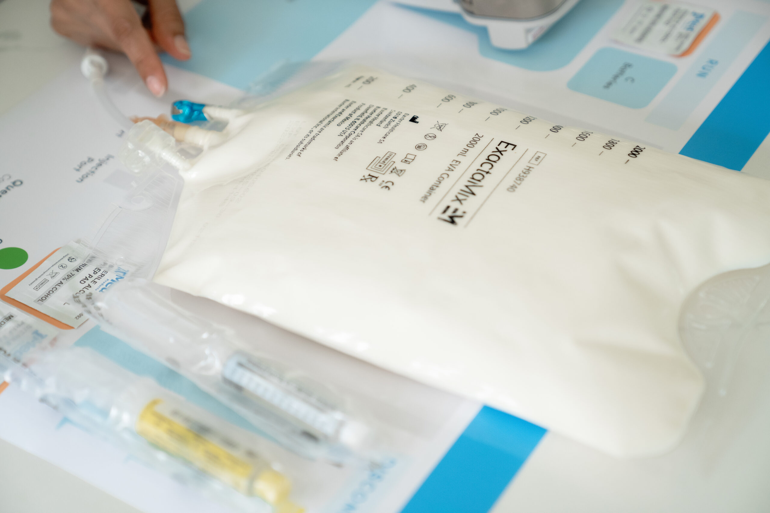
TPN, or total parenteral nutrition, is a method of intravenous nutrition delivery to patients, bypassing the gastrointestinal (GI) tract. As TPN is given intravenously, GI side effects such as diarrhea or stomach disturbances rarely occur.
However, if the treatment is administered incorrectly or without following necessary precautions or proper injection techniques, it can lead to infections at the injection site and other IV-related complications.
In this article, we will explore total parenteral nutrition’s side effects in detail and discuss ways to minimize them.
TPN provides vital nutrients directly into the bloodstream, bypassing the gastrointestinal tract. This approach is particularly beneficial for those unable to absorb nutrients through regular eating due to various medical conditions.
Learning about the mechanism of TPN helps you understand how side effects may arise. Here is an overview of what the treatment includes:
Intravenous Administration: TPN is administered through a special IV catheter, often a central venous catheter (CVC), giving direct access to the bloodstream.
Custom Nutrient Composition: The TPN mixture consists of macronutrients (carbohydrates, proteins, fats), micronutrients (vitamins, minerals), and electrolytes tailored to each individual’s needs.
Continuous or Cyclic Infusion: The solution can be delivered continuously or in cycles, depending on the patient’s health requirements and lifestyle.
Monitoring Parameters: Healthcare professionals monitor blood glucose, electrolyte levels, and weight to adjust the TPN formulation as necessary.
When the solution is improperly formulated or administered without adhering to protocols, it can lead to complications. Understanding TPN protocols allows healthcare providers to anticipate TPN’s side effects and implement preventative measures.
Side Effects of Total Parenteral Nutrition – Are They Serious?
All adverse effects of IV injections, such as TPN, can be serious and require immediate medical attention, regardless of whether they are common or rare. Because the medication enters the bloodstream directly, even minor issues may pose a serious, life-threatening risk.
TPN has a high sugar content, making it the perfect growth environment for bacteria. Hence, if the solution, tubing, or dressing gets contaminated or mishandled during or before administration, it can lead to bloodstream infections.
Infections can cause fever, chills, increased blood glucose levels, or a high white blood cell count, potentially resulting in severe complications that require prompt medical intervention.
Who Is at Risk for TPN’s Side Effects?
Some individuals are more susceptible to experiencing the adverse effects of TPN. Understanding these risk factors can help prioritize monitoring and care.
Patients receiving high concentrations of glucose in their TPN solutions
Categories of Total Parenteral Nutrition Side Effects
TPN’s side effects and complications can be divided into three categories based on their origin and impact on health:
Catheter-related problems
Metabolic problems (imbalances in nutrient levels or organ functions)
Sepsis (systemic infections resulting from contaminated IV solutions)
Let’s look closer at each of these categories below.
Catheter-Related Problems
Catheter-related problems are one of the top risks of TPN. These complications can occur due to a lack of aseptic and sterile practices, improper catheter flushing, and improper securement of the catheter. These complications can lead to thrombosis, phlebitis, systemic infections, and infections at the insertion site.
Metabolic Problems
Metabolic side effects can develop as a consequence of TPN treatment, affecting overall health. These imbalances often arise due to the specific nutrient composition of the TPN solution and the individual’s unique metabolic response.
Here are some of the most common metabolic issues patients may experience as side effects of total parenteral nutrition.
Hypoglycemia
One of the adverse effects of TPN is hypoglycemia or abnormally low blood sugar levels. It can happen during TPN therapy if glucose infusion occurs too rapidly or there is an inappropriate adjustment of insulin levels.
Symptoms may include dizziness, confusion, and fainting. If left unaddressed, severe hypoglycemia can lead to seizures, loss of consciousness, and potentially life-threatening complications.
Hyperglycemia
One of the other common side effects of TPN is hyperglycemia or elevated blood sugar levels. It often results from excessive glucose administration in TPN solutions.
Hyperglycemia may manifest as increased thirst, frequent urination, and fatigue. Without proper management, it can lead to dangerous complications such as diabetic ketoacidosis, long-term organ damage, and an increased risk of infections, particularly in those with diabetes.
Hepatic Dysfunction
Hepatic dysfunction refers to an impaired liver function that can develop due to prolonged TPN therapy, especially when there is an excess of calories or fat.
Hepatic side effects of TPN can manifest as elevated liver enzymes and jaundice. Without intervention, parenteral-associated hepatic dysfunction can progress to liver failure and necessitate more intensive treatments or even liver transplantation.
Metabolic Bone Disease
Metabolic bone disease encompasses a range of disorders resulting from imbalances in calcium, phosphorus, or vitamin D levels. These side effects of total parenteral nutrition more commonly occur in long-term TPN patients.
These conditions can weaken bones, increasing the risk of fractures and osteoporosis. If left untreated, bone diseases can severely impact mobility, causing chronic pain and disability.
Electrolyte Imbalance
The last type of TPN’s metabolic side effects is electrolyte imbalance or refeeding syndrome.
It can occur during TPN therapy, particularly if nutrition is restarted too quickly after a period of malnourishment and there are abnormal levels of essential minerals, such as sodium, potassium, or magnesium, in the body.
If not properly managed, these imbalances can cause serious complications, including cardiac arrhythmias, seizures, and muscle weakness.
Sepsis
Sepsis is a severe and potentially life-threatening condition that can result from infections linked to TPN administration. It occurs when bacteria enter the bloodstream, often through contaminated catheters or IV solutions.
Symptoms of sepsis may include fever, chills, rapid heart rate, and confusion. Immediate medical attention is critical to prevent severe outcomes and even death.
AmeriPharma® Specialty Care
Total Parenteral Nutrition | Leader In TPN Assistance
TPN’s complications can also be grouped by severity and how commonly they occur. Here is an overview of side effects based on these groupings.
Common Adverse Effects of TPN
The common side effects of total parenteral nutrition include:
Nausea or vomiting
Increased urination
Swelling of the hands, feet, or legs
Stomach pain
Tingling in the hands or feet
Fever or chills
Mouth sores
Skin changes
Rapid weight loss or gain
Muscle weakness
Severe Side Effects and Complications of TPN
Severe side effects are mostly catheter-related. These catheter-related problems are largely preventable through strict aseptic technique, including the proper handling of supplies, catheter access, as well as meticulous adherence to recommended TPN administration protocols. Failure to follow care instructions can increase the risk of catheter-related complications.
How to Prevent or Minimize TPN Side Effects and Complications
There are many ways to prevent the adverse effects of TPN. Here are some effective strategies to consider.
Aseptic Infusion Techniques
Follow aseptic infusion techniques, such as:
Ensure all equipment is clean and sanitized
Ensure supplies and TPN workspace are maintained in a clean area
Wash your hands thoroughly before handling any material
Wear gloves to minimize contamination risk during infusions, TPN setup, and connection
Supply packages should be opened only at the time of TPN preparation
Monitoring Patient Condition
Closely monitor the patient’s condition during TPN administration. Both patients and their caregivers must look for signs of infections at the injection site or other side effects while receiving TPN.
As directed, monitor the patient’s blood glucose levels, weight, and body temperature while on TPN therapy. Document any changes in symptoms and promptly report them to your healthcare team to facilitate appropriate interventions if needed.
Gradual Discontinuation Protocol
Do not stop total parenteral nutrition abruptly, as it can cause side effects such as hypoglycemia. Instead, develop a gradual weaning plan for a safe transition to alternative feeding methods.
Ensure regular blood glucose testing during this phase for timely adjustments and communicate closely with healthcare providers.
Controlled Infusion Rates
Administer TPN infusion at a slow and constant rate. Your Curlin pump will be pre-programmed to ensure it is not administered too rapidly, which can cause TPN-related side effects such as vomiting, nausea, or elevated body temperature.
The pharmacy team will monitor tolerance to TPN infusions. The infusion rate may be revised based on individual tolerance and response. This approach will ensure the recipient remains comfortable throughout the infusion process.
Catheter Management
To prevent catheter-related problems, always flush the catheter according to the healthcare provider’s instructions. Follow proper flush technique, including washing hands before and after touching flush syringes or the catheter.
To minimize infection risks, maintain proper dressing changes as performed by your nursing team. Regularly inspect the insertion site for signs of irritation or infection. Report any abnormal signs to your healthcare team in order to address issues proactively.
Long-Term Monitoring
Because bone or liver diseases can occur from long-term TPN use, the pharmacy team will monitor lab values during long-term therapy. Lab tests that are performed include liver function tests, blood levels of vitamin D, magnesium, phosphorus, and calcium.
Manage the Adverse Effects of TPN With AmeriPharma® Specialty Pharmacy
TPN’s side effects can be serious. Hence, if given at home, TPN must be administered and monitored by an experienced professional or trained person.
To avoid any side effects or complications of total parenteral nutrition, monitoring the injection site and overall patient’s condition is necessary, especially during the first 24 – 48 hours of TPN therapy.
At AmeriPharma® Specialty Pharmacy, we understand the unique challenges associated with TPN. Our specialized nursing team provides at-home TPN infusions, adapting to your schedule and helping you minimize the risk of TPN’s side effects.
Contact us today to speak to a specialist and receive specialty care with full-service coordination, copay assistance, and 24/7/365 support.
Hemophilia C is a rare blood disorder in which a clotting factor called factor XI is missing, low, or doesn’t work as it should. Because clotting factors are essential for clotting, having hemophilia C can put you at a higher risk of prolonged bleeding, especially after surgery, dental procedures, or injury.
Hemophilia C is a rare blood disorder. It affects about 1 in 100,000 people, according to the National Bleeding Disorders Foundation [1].
Hemophilia C can affect people of any age, sex, race, or ethnicity. Nonetheless, a higher prevalence — 1 in 450 individuals — has been reported in people of Ashkenazi Jewish descent [2].
2. How Is Hemophilia C Inherited?
Changes (mutations) in the gene that encodes clotting factor XI cause hemophilia C. Typically, these mutations pass from the parents to the child in an autosomal recessive pattern. This means an affected person gets the mutated gene from both parents.
The parents who carry one copy of the gene have partial factor XI deficiency. So they’re unlikely to have severe signs and symptoms of the condition.
Sometimes, a person may inherit the mutated gene in an autosomal dominant pattern. This means they may get hemophilia C even if only one parent has the condition.
3. What Is Acquired Hemophilia C?
Acquired factor XI deficiency is when a person gets the condition later in life. Unlike the inherited form, it doesn’t run in families.
The most common cause is liver disorder, which affects the production of factor XI. You may develop acquired hemophilia C if you’ve received a liver transplant from a factor XI-deficient donor.
In some cases, your immune system mistakenly produces proteins (autoantibodies) that attack factor XI, leading to factor XI deficiency.
4. What Are the Symptoms?
Not all people with a factor XI deficiency experience symptoms. Unlike other forms of hemophilia, lower levels of factor XI in the bloodstream don’t always result in more severe symptoms.
Symptoms can include:
Frequent nosebleeds
Easy bruising
Blood in urine
Unusual bleeding after dental procedures, surgery, or trauma
Heavy menstrual bleeding
Bleeding after childbirth
People with this condition rarely get joint and muscle bleeds.
5. What Is the Most Common Treatment for This Condition?
Treatment may not always be necessary, especially if bleeding isn’t problematic. However, those with mild symptoms may need to restrict activities that increase the risk of injury, such as contact sports.
You’ll usually need treatment before undergoing surgery or other procedures. In severe cases, you’ll need fresh frozen plasma (FFP), which is a clotting factor-rich blood derivative obtained from donors.
Other treatment options can include:
Antifibrinolytics, such as aminocaproic acid and tranexamic acid, to treat bleeding in the mouth and menstrual bleeding.
Medications to prevent the breakdown of replacement clotting factors.
Birth control pills for heavy menstrual bleeding
6. How Is Hemophilia C Treated During Pregnancy?
There are no evidence-based guidelines for hemophilia C treatment during pregnancy. The standard treatment is administering fresh frozen plasma at the time of delivery.
Treatment during pregnancy is anticipatory and requires a multidisciplinary care team.
Myasthenia gravis is a rare autoimmune disorder affecting 20 of every 100,000 people worldwide. There are various types of myasthenia gravis, each with slightly different symptoms and treatment options.
Whether you’re a person living with this disease or a family member caring for your loved one, knowing more about the type of myasthenia gravis you’re dealing with can help you navigate the treatment process.
Below, we will explore the different types of myasthenia gravis, their symptoms, and the available treatment options.
Myasthenia gravis (MG) is a chronic autoimmune disorder affecting the neuromuscular junction of the skeletal muscles. Various types of myasthenia gravis can target the muscles in your face, neck, eyes, arms, and legs, hindering your ability to:
Factors like genetics, infections, gender, and the presence of other autoimmune diseases may increase the risk of developing various types of myasthenia gravis. Once the symptoms appear, doctors will perform blood, imaging, and nerve function tests to diagnose the disease.
Types of Myasthenia Gravis
There are several different types of myasthenia gravis. Let’s examine each type and its symptoms to understand this disease better.
Generalized Myasthenia Gravis
Generalized myasthenia gravis, or gMG, is the most common form of the disease, accounting for 85% of all cases.
This form of the disease involves widespread muscle weakness that affects different areas. Some of gMG’s symptoms include:
Studies show that 55% of individuals with eye-related symptoms progress to developing generalized types of myasthenia gravis, usually within the first few years of symptom onset.
Approximately 10-40% of individuals with myasthenia gravis only experience eye-related symptoms due to the more vulnerable nature of the muscles in this area. This type of MG is called ocular myasthenia gravis, or OMG.
Symptoms of OMG include:
Eye strain
Blurred vision
Difficulty focusing
Diplopia (double vision)
Ptosis (drooping eyelids)
These symptoms may worsen with prolonged use of the eye muscles, such as when reading or watching TV. Ocular types of myasthenia gravis may later progress into gMG.
Seronegative Myasthenia Gravis
Seronegative MG is next on our list of different types of myasthenia gravis. Seronegative myasthenia gravis occurs when there are no identifiable autoantibodies (such as acetylcholine receptor antibodies) in the patient’s blood. Nearly 10% of MG cases are seronegative.
Symptoms of seronegative myasthenia gravis are similar to those of seropositive MG and include:
Double vision
Drooping eyelids
Difficulty speaking
Difficulty swallowing
Muscle fatigue and weakness
The absence of autoantibodies makes diagnosing the seronegative types of myasthenia gravis difficult. Patients may need to be evaluated by neurologists, and special tests may be performed to confirm seronegative MG.
Transient Neonatal Myasthenia Gravis
Transient neonatal myasthenia gravis (TNMG) occurs in 10-20% of babies born to affected mothers. Women who have myasthenia gravis during pregnancy or are in remission may pass their autoantibodies to the fetus.
Symptoms appear in the first 24 hours of the infant’s life and may include:
Weak crying
Muscle weakness
Respiratory problems
Lack of facial expressions
Difficulty suckling or swallowing
Babies with transient types of myasthenia gravis usually recover within a few weeks as the mother’s autoantibodies leave their bodies.
Congenital Myasthenia Gravis
Another one of the different types of myasthenia gravis is congenital MG or CMG. This form of MG stems from a genetic mutation and is inherited, unlike other acquired forms. It affects one in every 500,000 children from birth through early childhood.
Signs of CMG are similar to those of gMG and may include:
Facial weakness
Drooping eyelids
Muscle weakness
Breathing difficulties
Swallowing difficulties
However, symptoms may be milder in congenital types of myasthenia gravis if the disease appears in later stages of life.
Juvenile myasthenia gravis is a rare form of MG affecting 10-15% of children and teenagers with the condition. It usually begins before the patient turns 18 and lasts throughout their lives.
Symptoms of juvenile MG include:
Double vision
Drooping eyelids
Muscle weakness
Difficulty speaking
Difficulty breathing
Difficulty swallowing
Issues with fine motor skills
The overall symptoms of juvenile MG are similar to those in adults. However, they may appear differently in children and teenagers.
Late-Onset Myasthenia Gravis
The last form of the disease we cover is late-onset MG. Late-onset types of myasthenia gravis commonly occur in men over 50. They account for 15-20% of all cases with unique challenges.
Symptoms are similar to those of early-onset MG and may include:
Generalized fatigue
Chewing and swallowing difficulties
Drooping eyelids and vision problems
Muscle weakness (especially in the face and neck area)
While symptoms are similar to other types of MG, they may be less severe and progress more slowly.
Treatment Options for Different Types of Myasthenia Gravis
While we don’t yet have a cure, there are various treatment options for myasthenia gravis. Let’s explore these treatments and see which ones work best for various types of myasthenia gravis.
Medications
Certain medications can help alleviate the symptoms of myasthenia gravis. Two types of these medications include:
Cholinesterase inhibitors (anticholinesterase): These medications boost signals at the neuromuscular junctions, increasing acetylcholine levels and improving muscle strength.
Immunosuppressants: Drugs like corticosteroids suppress the immune system by reducing the production of harmful antibodies and preventing them from attacking the neuromuscular junctions.
Cholinesterase inhibitors are often prescribed for those with generalized, ocular, transient neonatal, and juvenile myasthenia gravis. Immunosuppressants are prescribed for the seronegative, congenital, and late-onset types of myasthenia gravis.
Monoclonal antibodies are another treatment option for different types of myasthenia gravis. They target specific cells and proteins in the immune system responsible for attacking the neuromuscular junction.
The most common monoclonal antibodies prescribed for MG are:
Rituximab: Targets B-cells, which play a crucial role in producing autoantibodies.
These monoclonal antibodies are typically prescribed for those with generalized, seronegative, ocular, and late-onset myasthenia gravis.
Plasma Exchange
Plasma exchange or plasmapheresis is an effective treatment for various types of myasthenia gravis. This method involves:
Removing the patient’s blood
Separating the plasma (containing the harmful autoantibodies) from other components
Returning the plasma to the patient’s body or replacing it with a substitute plasma solution
By removing the autoantibodies from the patient’s bloodstream, plasmapheresis can help alleviate symptoms. This treatment is usually prescribed for those with generalized, seronegative, late-onset, and severe cases of ocular myasthenia gravis.
IVIG
IVIG therapy is another popular treatment for different types of myasthenia gravis. This method involves infusing a concentrated solution of antibodies obtained from healthy donors into the patient’s bloodstream.
IVIG can be effective for generalized, seronegative, ocular, and juvenile types of myasthenia gravis.
Surgery
Surgery is the last treatment option for myasthenia gravis, reserved for cases where other therapies haven’t worked.
The goal of surgery is to remove the thymus gland, a gland located behind the sternum. Scientists believe that this gland is responsible for the production of autoantibodies. Therefore, the procedure is called a thymectomy.
This surgery may be suitable for younger patients with generalized and seronegative MG.
Living With Myasthenia Gravis
Living with myasthenia gravis can be challenging. However, there are things you can do to make life with myasthenia gravis easier.
Here are some tips to deal with the daily challenges of various types of myasthenia gravis:
Use mobility aids and assistive devices when necessary
Get plenty of rest and listen to your body to conserve energy
Eat a balanced diet and stay hydrated to support your overall health
Stay active with gentle exercises like yoga to maintain muscle strength
Lean on friends and family for emotional support and practical assistance
Take your medications as prescribed and report any changes to your doctor
Join a support group for individuals with myasthenia gravis to connect with others
Practice relaxation techniques like meditation and deep breathing to manage stress
Avoid triggers like extreme temperatures or overexertion to keep the symptoms from worsening
Receive At-Home Myasthenia Gravis Treatment from AmeriPharma® Specialty Pharmacy
Now that you know more about the various types of myasthenia gravis, you are prepared to face this disease head-on and overcome any challenges that may arise.
Our URAC and ACHC-accredited specialty pharmacy offers at-home IVIG therapy to individuals battling myasthenia gravis in 40+ US states and territories. Our trained infusion nurses can come to your home to administer the treatment and offer troubleshooting support.
Contact us now to receive specialty care at home and manage your condition with full-service coordination, 24/7/365 support, and copay assistance.
Autoimmune encephalitis (AIE) is a rare and complex neurological condition with symptoms that can range from mild confusion to coma. Due to its severity, those affected usually require intensive care over long periods of time.
Fortunately, there are several long-term treatment options for autoimmune encephalitis. Here, we will explore five autoimmune encephalitis treatments that have been proven effective by studies and patients.
Let’s start with a quick guide to autoimmune encephalitis. In this rare autoimmune disorder, the body’s immune system attacks the brain, causing inflammation and symptoms such as:
Seizures
Insomnia
Numbness
Panic attacks
Hallucinations
Severe anxiety
Impaired memory
Cognitive difficulties
Compulsive behaviors
Involuntary movements
Difficulty with balance and speech
Symptoms may vary in different subtypes of the disease. However, if left untreated, AIE can become dangerous and even lead to severe coma.
What Causes Autoimmune Encephalitis?
AIE affects around 13 out of every 100,000 individuals. You might wonder, since we have developed effective autoimmune encephalitis treatments, we must know what exactly causes this disease. However, that is not the case in many instances.
While we may not know the exact cause of the disease in all cases, scientists believe it may be triggered by:
Certain types of cancers
Presence of a tumor called teratoma
Exposure to certain bacteria and viruses
How Is Autoimmune Encephalitis Diagnosed?
Diagnosing this condition can be difficult. Sometimes, doctors mistake it for other neurological conditions and even drug abuse.
However, if you visit a neurologist with signs of AIE, they will perform the following tests to confirm the disease:
MRI
Blood tests
A spinal tap
Neurologic exams
Effective Autoimmune Encephalitis Treatments
After diagnosis, your doctor can decide which long-term treatment for autoimmune encephalitis is best for you. Here are five of the most effective treatments for AIE.
Intravenous immunoglobulin (IVIG) is a first-line therapy for autoimmune encephalitis. IVIG infusion treatment involves administering a concentrated dose of antibodies extracted from healthy donated blood plasma to AIE patients.
IVIG’s mechanism of action relies on these antibodies. They neutralize autoantibodies that are causing the disease and regulate the immune response to alleviate symptoms. According to studies, IVIG is one of the safest and most effective treatments for autoimmune encephalitis.
Patients undergo a thorough evaluation by a neurologist.
If IVIG is recommended, the physician will schedule a series of infusions.
IVIG can be administered in a hospital, an outpatient infusion center, or at home, under the supervision of a healthcare professional.
A trained nurse will administer the prepared solution through an IV line.
They will monitor the recipient’s vital signs for any adverse reactions.
The treatment might take anywhere from 1 hour to 4 hours.
Afterward, patients might feel a slight headache and fever that will subside quickly.
Patients may require follow-up appointments with their providers to assess the effectiveness of the autoimmune encephalitis treatment.
2. Plasmapheresis
Another long-term treatment for autoimmune encephalitis is plasmapheresis (plasma exchange). This technique involves removing and replacing the person’s plasma to eliminate harmful antibodies and inflammatory substances.
Plasmapheresis results in the rapid improvement of symptoms, which can be helpful for patients with severe refractory AIE.
Plasmapheresis Administration for Autoimmune Encephalitis
Administering plasmapheresis to AIE patients involves the following steps:
The doctor thoroughly evaluates the individual to determine if plasma exchange is the best treatment for them.
Patients may need to fast before receiving this autoimmune encephalitis treatment. They may also need to take medications to minimize the side effects of treatment.
Trained medical professionals typically administer this treatment in a hospital setting.
They will insert a central venous catheter into a large vein in the arm, neck, or groin.
This catheter will remove the blood and circulate it through a machine to separate the plasma.
The plasma with harmful antibodies is filtered, and healthy plasma is returned to the individual’s bloodstream.
Once the desired plasma volume has been exchanged, the doctors will remove the catheter and allow the patient to rest while monitoring their vital signs.
3. Rituximab and Other Monoclonal Antibodies
Rituximab and monoclonal antibodies are among the most effective autoimmune encephalitis treatments. These targeted therapies are especially effective for patients who have not responded to other forms of treatment.
These monoclonal antibodies work by targeting specific immune system components (such as B cells) to control autoimmune processes in the brain.
Rituximab Administration for Autoimmune Encephalitis
Administering rituximab and other monoclonal antibodies as long-term treatments for autoimmune encephalitis includes the following steps:
A healthcare provider will evaluate the patient to determine the appropriateness of targeted immunotherapy.
They will determine the dosage of rituximab and the treatment schedule based on the patient’s weight and AIE subtype.
The medication is typically infused in cycles over several weeks or months.
The patient is closely monitored throughout autoimmune encephalitis treatment for side effects and disease progression.
Patients will attend follow-up appointments to assess their response to medication and adjust the treatment plan as needed.
Corticosteroids such as prednisone and methylprednisolone are effective treatment options when used in combination with other therapies.
These medications work by reducing inflammation in the brain and suppressing the immune response. This action alleviates neurological symptoms in affected individuals and improves their quality of life.
Corticosteroids Administration for Autoimmune Encephalitis
Here is how corticosteroids are administered to AIE patients:
Before starting this treatment for autoimmune encephalitis, patients undergo a comprehensive evaluation by a physician.
The physician will determine the appropriate dosage and duration of therapy based on clinical symptoms.
They will prescribe a specific corticosteroid medication based on the patient’s needs.
The patient will receive treatment orally in pill form or intravenously through an IV drip.
The physician will monitor the patient’s response to treatment and adjust the dosage and duration of therapy if necessary.
As the patient’s symptoms improve, the physician will gradually reduce the dosage and monitor the patient for relapse.
5. Stereotactic Radiosurgery
The last effective autoimmune encephalitis treatment on our list is stereotactic radiosurgery. This long-term treatment for autoimmune encephalitis is a non-invasive procedure. It uses high doses of radiation to target specific areas in the brain.
This treatment is best for patients with focal brain regions resistant to other therapies. The radiation will reduce inflammation and control autoimmune processes without the need for open surgery.
Stereotactic Radiosurgery Operation for Autoimmune Encephalitis
The procedure is delivered to AIE patients in the following steps:
Affected individuals undergo MRI or CT scans to identify target lesions.
An interdisciplinary team of neurologists, neurosurgeons, and radiation specialists collaborate to determine whether radiosurgery is a suitable treatment for the person with autoimmune encephalitis.
Based on the results, they develop a treatment plan for the patient.
They use computer algorithms and specialized software to map the exact locations of lesions and spare the surrounding healthy areas.
For the surgery, patients lie down on a treatment table, and the radiation specialists deliver the beams to the identified areas.
Patients may require multiple sessions.
Afterward, they are monitored for side effects and changes in symptoms.
Challenges in Treating Autoimmune Encephalitis
While there have been advancements in the treatment of autoimmune encephalitis, several challenges still exist. Let’s look at some of them.
One of the biggest challenges in administering long-term treatment for autoimmune encephalitis is delays in diagnosis.
Due to the similarity of AIE’s symptoms to other neurological disorders, doctors may misdiagnose the condition. This delayed diagnosis will lead to the progression of symptoms and delays in receiving appropriate treatment.
Side Effects
Some patients may experience severe side effects with current treatments. These side effects may include:
Infections
Allergic reactions
Damage to healthy tissues
Treatment Resistance
In some cases, patients may develop resistance to autoimmune encephalitis treatments. The medications will no longer work, and the condition becomes unmanageable.
This resistance can complicate treatment decisions, necessitating a more personalized approach.
Access to Specialized Care and Medications
The last challenge in treating autoimmune encephalitis is the lack of access to specialized care and medications. Many patients live in areas where they do not have access to specialized healthcare providers.
No neurologists or immunologists are available in their vicinity to make a correct diagnosis. Additionally, they may not have access to specialized medications used in treating AIE.
These are some of the primary challenges individuals with autoimmune encephalitis face. By addressing these issues and developing more targeted treatments, we can enhance the quality of care for those living with this debilitating neurological disorder.
Fight Autoimmune Encephalitis With AmeriPharma® Specialty Pharmacy
Autoimmune encephalitis is not an unbeatable disease. If your doctor diagnoses it early, you can receive effective autoimmune encephalitis treatment and manage your condition.
If you are looking for accessible long-term treatments for autoimmune encephalitis, AmeriPharma® Specialty Pharmacy can help.
We offer at-home IVIG therapy to those battling this autoimmune disorder in 40+ US states and territories. Our specialty pharmacy can even send specialized infusion nurses to your home to administer the treatment and help you troubleshoot any problems.
Contact us now to receive specialty care at home and manage AIE with our full-service coordination, 24/7/365 support, and copay assistance.
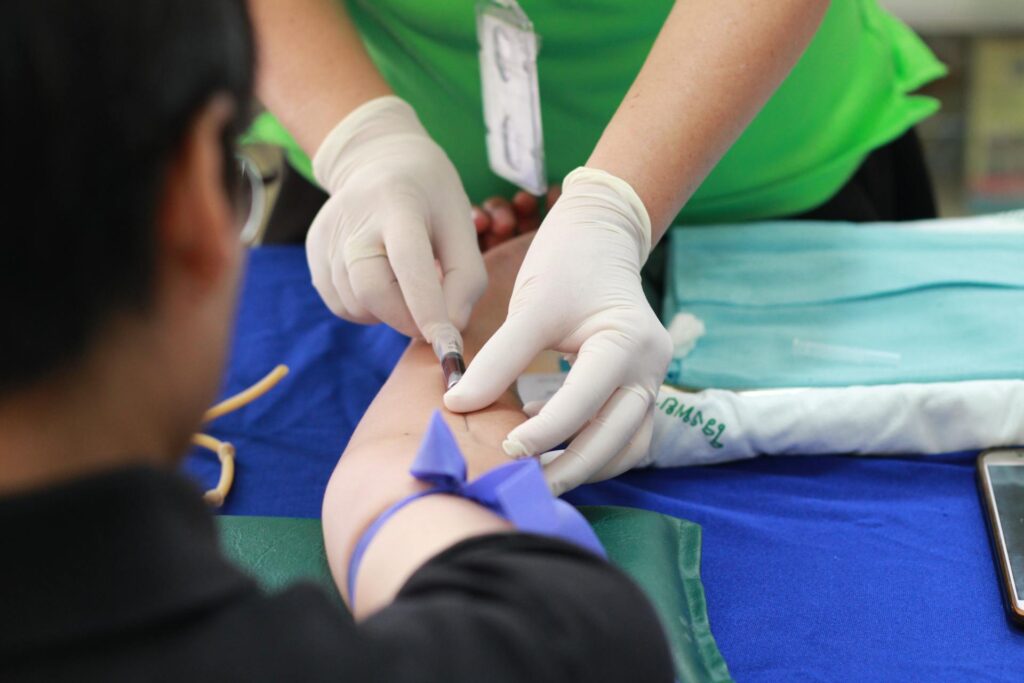
IVIG is used as a supportive therapy in patients with multiple myeloma. Multiple myeloma is a rare type of blood cancer that forms in plasma cells, a type of white blood cell that produces antibodies to fight infections under normal conditions.
In patients with multiple myeloma, the cancerous plasma cells do not produce effective antibodies, leaving them more vulnerable to infections. According to research, recurrent infections are one of the underlying causes of death in 22% to 45% of patients with multiple myeloma.
Doctors typically recommend IVIG therapy to reduce mortality and morbidity rates in multiple myeloma patients.
Let’s review the basics of multiple myeloma, including its effects on the immune system, as well as how IVIG therapy can help patients suffering from multiple myeloma.
Basics of Multiple Myeloma and How It Affects the Immune System
Multiple myeloma is a cancer that arises from the bone marrow and affects plasma cells. It is also known as“cancer of plasma cells,” where a single plasma cell becomes cancerous, multiplies nonstop, and builds up in the bone marrow. Eventually, the cancerous plasma cells (also known as myeloma cells) crowd out the healthy blood-forming cells in the bone marrow.
Furthermore, cancerous plasma cells produce abnormal, ineffective antibodies called M proteins that can’t fight infections.
Additionally, myeloma treatments like chemotherapy and stem cell transplants weaken the patient’s immune system. These therapies not only kill the cancerous cells but also destroy the healthy cells, leading to low healthy plasma cell counts and antibody levels. As a result, patients with multiple myeloma become highly susceptible to infections such as pneumonia (respiratory infection).
What Is the Prevalence of Multiple Myeloma in the US?
According to American Cancer Society estimates, over 35,000 new cases of multiple myeloma and around 12,540 deaths are expected to occur in 2024.
Multiple myeloma is more common in older people, with an average age of 65 – 69 years old.
How Can IVIG Therapy Help Multiple Myeloma Patients?
IVIG therapy has been used for decades to treat various immune-related conditions, including multiple myeloma.
In multiple myeloma, IVIG therapy temporarily supplies patients with an array of antibodies and helps to improve their overall immune functions. These antibodies recognize and fight off infections that the patient’s own immune system is too compromised to handle.
How Does IVIG Therapy Work?
Although the exact mode of action of IVIG therapy is unknown, according to studies, it boosts the immune response against infections. In multiple myeloma patients, IVIG can exert the following beneficial effects:
It Reduces the Frequency of Recurrent Infections
IVIG therapy strengthens the patient’s immune system by providing antibodies. Various studies indicate that IVIG therapy can help reduce the frequency of recurrent infections, including grade 3 infections, in patients with multiple myeloma.
For example, a study published in ”Blood Cancer Discovery” reported that intravenous immunoglobulin therapy can reduce the risk of grade 3 and grade 5 infections by 90% in patients with relapsed or refractory multiple myeloma. Researchers further concluded that IVIG therapy should be given throughout treatment as well as after treatment.
IVIG provides passive immunity to MM patients, which means ready-made antibodies obtained from donors are infused into the body of an MM patient to fight infections. Unlike vaccines that stimulate the plasma cells to produce antibodies, IVIG therapy supplements the patient’s body with essential antibodies that provide immediate protection and work against infections.
It Exerts Anti-Cancer and Anti-Inflammatory Effects
IVIG is known for its anti-inflammatory properties, which help to regulate the overactive immune system in various autoimmune disorders.
Recent research at the MD Anderson Cancer Center revealed that IVIG therapy decreased the growth of myeloma cells (cancerous plasma cells) and caused cell death. Based on these findings, the researcher concluded that IVIG therapy may induce anti-cancer effects and that adding it to standard myeloma treatment could help obtain better outcomes.
What Is the Standard Dose of IVIG for Multiple Myeloma Patients?
IVIG is typically given to myeloma patients every 3 to 4 weeks at a dosage of 0.4 grams per kilogram. The frequency of IVIG administration may differ depending on the person’s steady-state immunoglobulin levels.
For example, some patients may require IVIG therapy once a month, while others may need it more frequently.
Who Can Benefit From IVIG Therapy?
Multiple myeloma patients who have hypogammaglobulinemia (low level of antibodies in their blood) and are highly susceptible to recurrent or severe infections can benefit from IVIG therapy.
Similarly, MM patients who are undergoing chemotherapy and stem cell transplants can also get benefits from IVIG therapy, as these intensive treatments affect the immune system and increase the risk of infections.
It is important to note that IVIG therapy is not a stand-alone treatment for multiple myeloma. Rather it is used as a supportive therapy to reduce the risk of infections in multiple myeloma patients.
Hemophilia is a rare genetic bleeding disorder that predominantly affects men, though it can also occur in women. Historically, hemophilia has been considered a “man’s disease,” leading to the widespread misconception that women could only be carriers — passing the gene to their children — without being affected themselves. However, this is not the case.
Women can also have hemophilia, but it manifests differently than in men. In fact, according to the CDC’s Community Counts HTC data, more than 2,700 women in the U.S. have hemophilia A or B.
This article discusses the genetic background and conditions under which women can develop hemophilia.
Hemophilia is an X-linked disorder, which means the mutated genes that code for clotting factor proteins (factor VIII and factor IX) are present on the X chromosomes.
Genetically, males have one X chromosome (inherited from their mother) and a Y chromosome (received from their father). When a male gets the affected X chromosome (which has the faulty gene) from a carrier mother, he develops hemophilia due to a deficiency of clotting factors.
In contrast, females have two X chromosomes, which they inherit from each parent. The normal gene on one chromosome overrides the mutated gene on the affected chromosome. For this reason, women are the “carriers” of hemophilia, which means they carry defective genes but do not develop hemophilia. This is why hemophilia is more commonly observed in men as men only have one copy of the X chromosome.
However, carrier women may experience some hemophilia symptoms. For example, about 1 out of 4 carrier women experience mild bleeding symptoms such as nosebleeds or heavy menstrual periods due to low clotting factor levels.
What Are the Conditions That Causes Hemophilia in Women?
Women can have hemophilia under the following two conditions:
1. Homozygous Condition
In the homozygous condition, when the daughter gets two affected chromosomes from each parent, she develops hemophilia. However, this condition is extremely rare and can only happen if a father has hemophilia and a mother is a carrier.
2. Skewed X Chromosome Inactivation
In skewed X chromosome inactivation, when a woman receives one affected chromosome, and the other X chromosome with the normal copy of the genes is not working properly or “turned off,” she develops hemophilia due to the deficiency of clotting factors. This can happen for various genetic reasons.
Women with one or more affected genes can experience symptoms like men. Depending on the clotting factor levels in their blood, symptoms can range from mild to severe.
Here are some of the common symptoms that hemophilic women may experience:
Heavy bleeding from dental work, surgeries, or childbirth
Excessive and frequent nosebleeds that last longer than 10 minutes
Joint damage
Easy bruising
In addition to the above, hemophilic women often experience heavy menstrual bleeding (menorrhagia). Prolonged menstrual bleeding can cause anemia and iron deficiency.
The signs of heavy menstrual bleeding include:
Bleeding for more than 7 days
Passing clots that are bigger than a bottle cap
Changing a tampon or pad every 2 hours or less
Flooding or gushing of blood
If you or your loved one experiences any of the above symptoms (especially menorrhagia), immediately consult your healthcare provider for the diagnosis of hemophilia.
Conclusion
Although hemophilia is more frequently diagnosed in men, women can also have hemophilia; however, these instances are frequently underrecognized or misunderstood. Due to conditions like X-chromosome inactivation or having two faulty X chromosomes, women who carry the hemophilia gene may develop mild to severe symptoms. Timely diagnosis and efficient treatment can help to improve the quality of life in women with hemophilia.